. 2014 Jun 1;134(11):2633-45.
doi: 10.1002/ijc.28590. Epub 2014 Jan 20.

Origin and pharmacological modulation of tumor-associated regulatory dendritic cells

Affiliations

Origin and pharmacological modulation of tumor-associated regulatory dendritic cells

Hua Zhong et al. Int J Cancer. .

Abstract

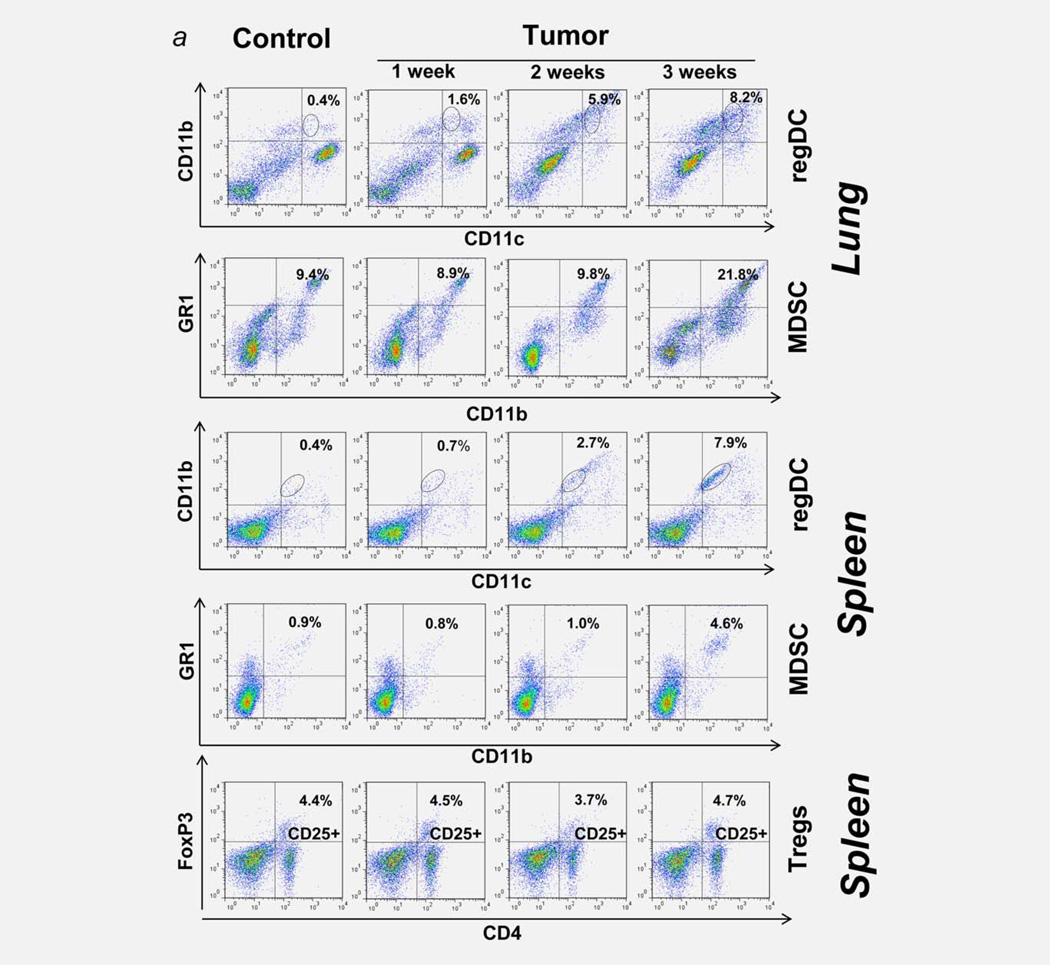
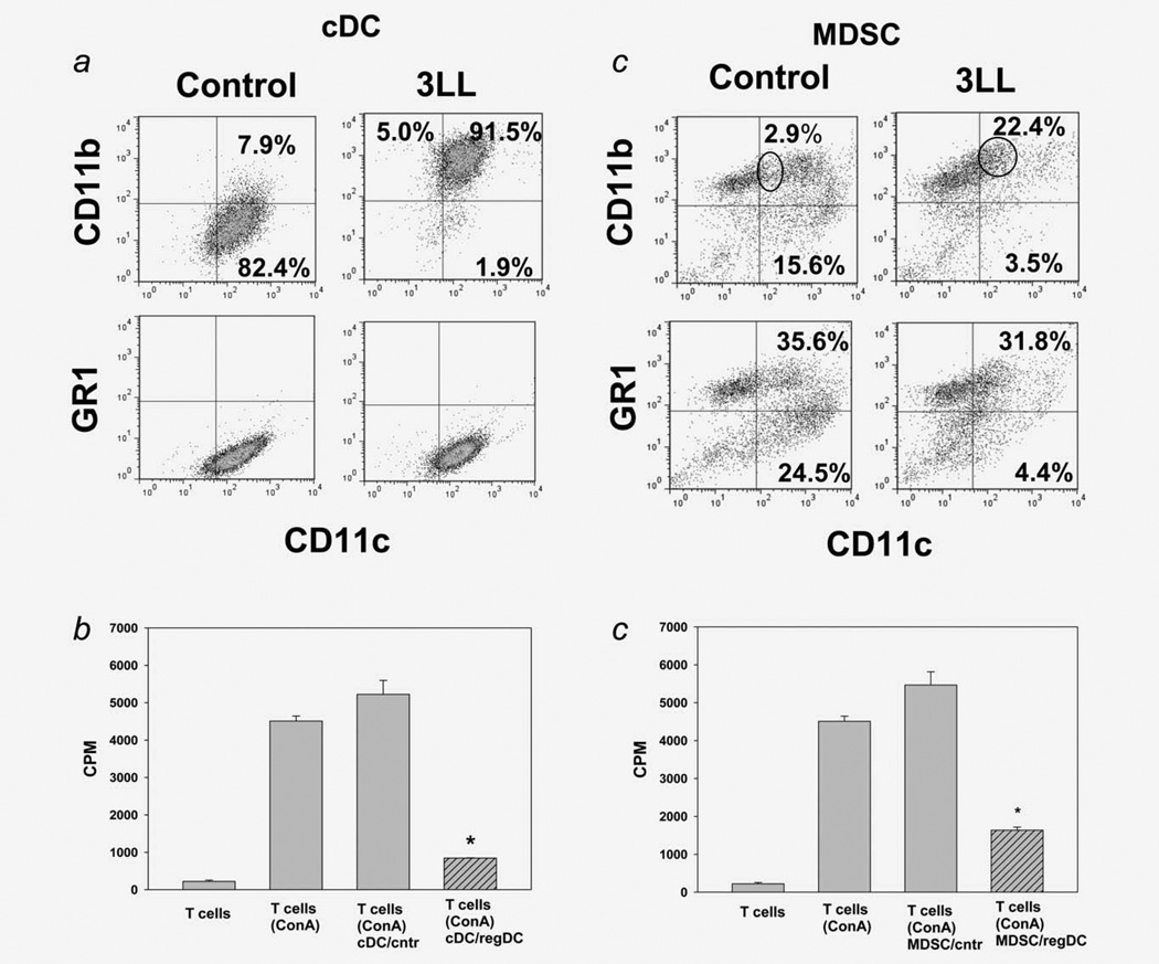
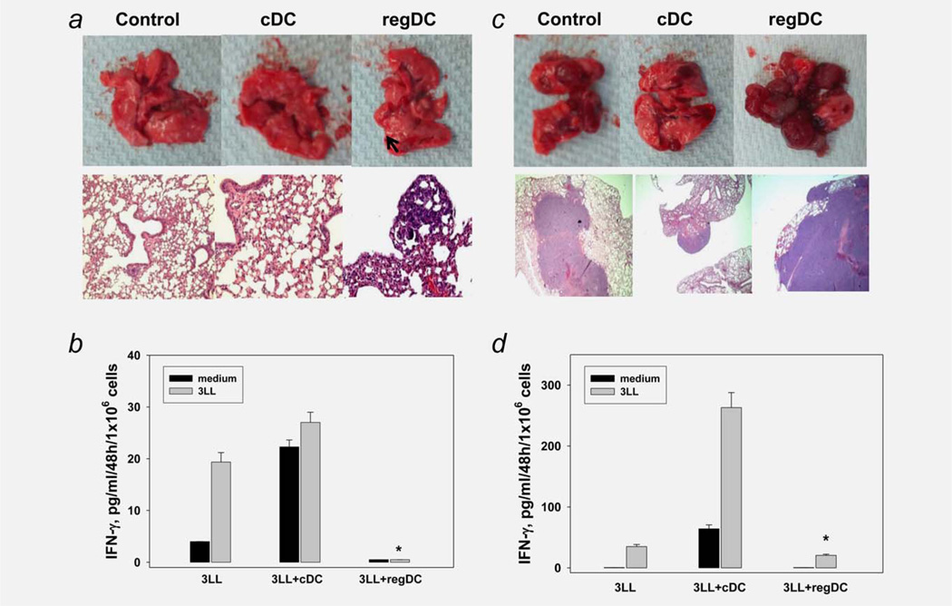
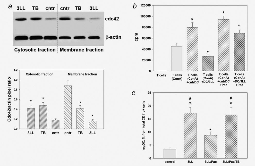
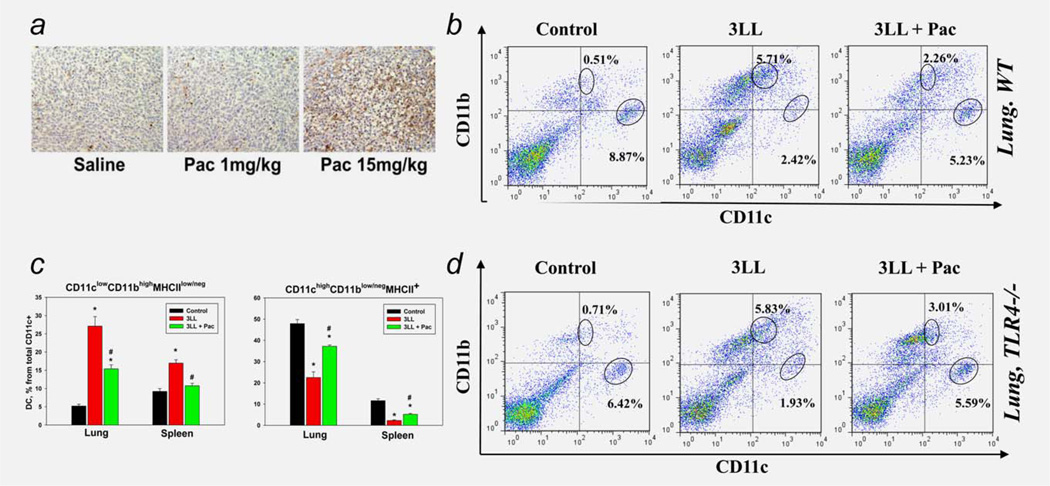
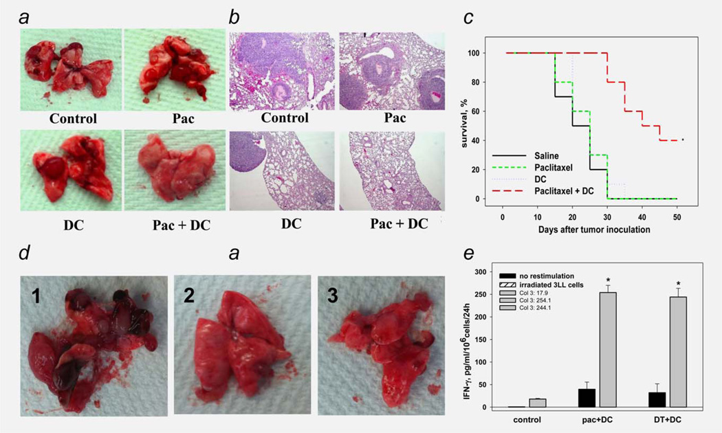
Protumorigenic activity of immune regulatory cells has been proven to play a major role in precluding immunosurveillance and limiting the efficacy of anticancer therapies. Although several approaches have been offered to deplete myeloid-derived suppressor cells (MDSC) and regulatory T cells, there are no data on how to control suppressive dendritic cell (DC) accumulation or function in the tumor environment. Although immunosuppressive function of DC in cancer was implicated to immature and plasmacytoid DC, details of how conventional DC (cDC) develop immunosuppressive properties remain less understood. Here, we show that the development of lung cancer in mice was associated with fast accumulation of regulatory DC (regDC) prior to the appearance of MDSC. Using the in vitro and in vivo approaches, we demonstrated that (i)both cDC and MDSC could be polarized into protumor regDC in the lung cancer environment; (ii) cDC → regDC polarization was mediated by the small Rho GTPase signaling, which could be controlled by noncytotoxic doses of paclitaxel; and (iii) prevention of regDC appearance increased the antitumor potential of DC vaccine in lung cancer. These findings not only bring new players to the family of myeloid regulatory cells and provide new targets for cancer therapy, but offer novel insights into the immunomodulatory capacity of chemotherapeutic agents used in low, noncytotoxic doses.

Keywords: MDSC; dendritic cells; small Rho GTPases; taxol; tumor microenvironment.


Figures

Figure 1
Figure 1
Immune regulators in the lung cancer microenvironment. 3LL-induced polarization of cDC into immunosuppressive regDC in vivo. (a) Early emergence of regDC, but not MDSC or Treg cells, in the lung and spleen after tumor cell inoculation. Mice received 3LL cells and tissues were collected 7, 14 and 21 days later as described in M&M. Flow cytometry analysis of CD11clowCD11bhigh/med regDC, MDSC and Treg cells revealed appearance of regDC within the first week after tumor cell injection, while the levels of MDSC and Treg cells increased 2 weeks later. Data are shown as the percentage of positive cells from a representative experiment (N=4–5). (b) Low expression of co-stimulatory molecules on tumor-associated regDC. Lungs were harvested from tumor-free (left panel) and 3LL-bearing (right panel) mice. Single cell suspensions were subjected to flow cytometric analysis as described in M&M. Significant upregulation of CD11clowCD11bhigh regDC and downregulation of CD11chighCD11blow/neg cDC were seen in all experiments (N>10). Data are shown as the percentage of positive cells from a representative experiment. (c) Polarization of bone marrow-derived cDC into immunosuppressive regDC in the lung cancer microenvironment in vitro. DC were generated from the bone marrow precursors and treated with 3LL-conditioned medium on Day 5 as described in M&M. Flow cytometry analysis revealed increased levels of regDC and decreased content of cDC subsets. The results are shown as the percentage of positive cells in a representative experiment. Similar data were obtained in seven independent studies. (d) Immunosuppressive activity of tumor-associated regDC. CD11clowCD11bhigh regDC were sorted from the lungs harvested 2 weeks after mice were injected with 3LL cells (regDC/3LL in vivo) or from the bone marrow-derived DC treated on Day 5 with 3LL-conditioned medium for 48 hr (regDC/3LL in vitro). Control CD11chighCD11blow/neg cDC were either bone marrow-derived cDC or lung cDC isolated from tumor-free mice (cDC). All DC subsets were added at the same ratio (1:100) to ConA-prestimulated syngeneic T cells. T cell proliferation was assessed by 3H-thymidine incorporation and expressed as count per minute (cpm). Proliferation of intact T cells and ConA-stimulated T cells (w/o DC) is shown as white bars. *p <0.05 (ANOVA, mean ± SEM, N = 5–6). (e) Inability of immature DC (imDC) to suppress proliferation of T lymphocytes. Bone marrow-derived imDC and conventional cDC were generated as described in M&M. All DC subsets were added at the same ratio to ConA-prestimulated syngeneic T cells. T cell proliferation was assessed by 3H-thymidine incorporation. Proliferation of intact T cells and ConA-stimulated T cells (w/o DC) is shown as white bars. *p <0.05 (ANOVA, mean ± SEM, N = 3). [Color figure can be viewed in the online issue, which is available at wileyonlinelibrary.com.]
Figure 1
Figure 1
Immune regulators in the lung cancer microenvironment. 3LL-induced polarization of cDC into immunosuppressive regDC in vivo. (a) Early emergence of regDC, but not MDSC or Treg cells, in the lung and spleen after tumor cell inoculation. Mice received 3LL cells and tissues were collected 7, 14 and 21 days later as described in M&M. Flow cytometry analysis of CD11clowCD11bhigh/med regDC, MDSC and Treg cells revealed appearance of regDC within the first week after tumor cell injection, while the levels of MDSC and Treg cells increased 2 weeks later. Data are shown as the percentage of positive cells from a representative experiment (N=4–5). (b) Low expression of co-stimulatory molecules on tumor-associated regDC. Lungs were harvested from tumor-free (left panel) and 3LL-bearing (right panel) mice. Single cell suspensions were subjected to flow cytometric analysis as described in M&M. Significant upregulation of CD11clowCD11bhigh regDC and downregulation of CD11chighCD11blow/neg cDC were seen in all experiments (N>10). Data are shown as the percentage of positive cells from a representative experiment. (c) Polarization of bone marrow-derived cDC into immunosuppressive regDC in the lung cancer microenvironment in vitro. DC were generated from the bone marrow precursors and treated with 3LL-conditioned medium on Day 5 as described in M&M. Flow cytometry analysis revealed increased levels of regDC and decreased content of cDC subsets. The results are shown as the percentage of positive cells in a representative experiment. Similar data were obtained in seven independent studies. (d) Immunosuppressive activity of tumor-associated regDC. CD11clowCD11bhigh regDC were sorted from the lungs harvested 2 weeks after mice were injected with 3LL cells (regDC/3LL in vivo) or from the bone marrow-derived DC treated on Day 5 with 3LL-conditioned medium for 48 hr (regDC/3LL in vitro). Control CD11chighCD11blow/neg cDC were either bone marrow-derived cDC or lung cDC isolated from tumor-free mice (cDC). All DC subsets were added at the same ratio (1:100) to ConA-prestimulated syngeneic T cells. T cell proliferation was assessed by 3H-thymidine incorporation and expressed as count per minute (cpm). Proliferation of intact T cells and ConA-stimulated T cells (w/o DC) is shown as white bars. *p <0.05 (ANOVA, mean ± SEM, N = 5–6). (e) Inability of immature DC (imDC) to suppress proliferation of T lymphocytes. Bone marrow-derived imDC and conventional cDC were generated as described in M&M. All DC subsets were added at the same ratio to ConA-prestimulated syngeneic T cells. T cell proliferation was assessed by 3H-thymidine incorporation. Proliferation of intact T cells and ConA-stimulated T cells (w/o DC) is shown as white bars. *p <0.05 (ANOVA, mean ± SEM, N = 3). [Color figure can be viewed in the online issue, which is available at wileyonlinelibrary.com.]
Figure 2
Figure 2
Tumor-induced polarization of cDC and MDSC into regDC ex vivo. (a) CD11c+ cDC and MDSC were isolated by FACS cell sorting from the lungs of tumor-free mice and used for phenotypic and functional assays. (b) Polarization of cDC into regDC ex vivo. Lung CD11c+ cDC were cultured ex vivo with 3LL cells in inserts for 72 hr. Flow cytometric analysis revealed the appearance of GR-1 negative regDC. The results are shown as the percentage of positive cells in a representative experiment with similar results obtained from four independent experiments. (c) Inhibition of T cell proliferation by cDC-derived regDC. Control lung CD11c+cDC or cDC-derived regDC were harvested and added at the same ratio (1:100) to ConA-prestimulated syngeneic T cells. T cell proliferation was assessed by 3H-thymidine incorporation and expressed as count per minute (cpm). *p <0.05 (ANOVA, mean ± SEM, N = 3). (d) Polarization of MDSC into regDC ex vivo. MDSC isolated from lung of tumor free mice were cultured ex vivo with 3LL cells in inserts for 72 hr. Tumor cells polarized lung MDSC into the regDC ex vivo. (e) Inhibition of T cell proliferation by MDSC-derived regDC. Tumor-induced regDC derived from MDSC were isolated by FACS cell sorting from the cell cultures and used in T cell proliferation assay as described in (c). MDSC-derived regDC significantly inhibited proliferation of preactivated T cells as compared to control untreated MDSC. *p <0.05 (ANOVA, mean ± SEM, N = 3).
Figure 3
Figure 3
Regulatory DC support tumor growth and attenuate antitumor immunity. (a) Administration of regDC but not cDC induced tumor development in the lung. Sorted in vitro generated regDC and cDC were injected in mice on Days 3 and 10 after inoculation of 1/10 of the minimum tumorigenic dose of 3LL cells. Animals were sacrificed at Day 21 and the lungs were removed, fixed and subjected to H&E staining. Control animals received only 3LL cells. Upper panels: visual appearance of tumor nodules in the lungs after administration of regDC. Low panels: H&E staining of the lung tissues for visualization of micronodules of lung carcinoma. The results of a representative experiment are shown (N = 3). (b) Inhibition of tumor-specific CTL by regDC in vivo. Splenic T lymphocytes were isolated from mice receiving either 1/ 10 minimum tumorigenic dose of tumor cells only (3LL), tumor cells and cDC (3LL + cDC) or tumor cells and regDC (3LL + regDC). Cells were stimulated with medium (black bars) or irradiated 3LL cells (gray bars). IFN-γ levels in cell-free supernatants were assessed by ELISA. Data represent the mean ± SEM from three independent experiments. * p <0.05 vs. 3LL and 3LL + cDC groups (ANOVA). (c) Administration of regDC, but not cDC augmented tumor development in the lungs. Sorted regDC and cDC were injected in mice together with 3LL cells at the minimum tumorigenic dose and the lungs were harvested 3 weeks after tumor cell injection. Upper panels: visual appearance of tumor nodules in the lungs. Low panels: H&E staining of the lung tissues for histopathological visualization of micronodules of lung carcinoma (×100). The results of a representative experiment are shown (N = 3). (d) Inhibition of tumor-specific CTL by regDC in vivo. Splenic T lymphocytes were isolated from mice receiving either tumor cells only (3LL), tumor cells and cDC (3LL + cDC) or tumor cells and regDC (3LL + regDC). Cells were stimulated with medium (black bars) or irradiated 3LL cells (gray bars). IFN-γ levels in cell-free supernatants were assessed by ELISA. Data represent the mean ± SEM from three independent experiments. *p <0.05 vs. 3LL and 3LL + cDC groups (ANOVA).
Figure 4
Figure 4
Regulation of tumor-induced formation of regDC by Rho GTPases and paclitaxel in vitro. (a) Decrease in active (membrane) and increase in inactive (cytosolic) Cdc42 in 3LL-treated DC. Bone marrow-derived DC were treated with medium (cntr), small Rho GTPase inhibitor toxin B (TB) and 3LL conditioned medium (3LL). Active and nonactive Cdc42 was assessed as Cdc42 protein levels in the membrane and cytosolic fractions of DC, respectively, by Western blot analysis (upper panel) as described in M&M. The results of a representative experiment are shown (N = 3). Densitometry was used for enumeration of the protein redistribution data (low panel). Data are shown as the mean ± SEM of Cdc42 levels relative to β-actin levels in the same fractions. *p <0.05 vs. control (ANOVA, N = 3). (b) Paclitaxel prevented 3LL-induced polarization of cDC into regDC in vitro. Bone marrow-derived cDC were polarized into regDC by the addition of 3LL conditioned medium (3LL) in the presence or absence of paclitaxel (Pac, 1 nM). Functional activity of medium-treated DC (cntrDC) and 3LLtreated cells (DC/3LL) was assessed by their ability to suppress proliferation of ConA-preactivated syngeneic T cells. T cell proliferation was determined by 3H-thymidine incorporation and expressed as count per minute (cpm). Proliferation of intact T cells and ConA-stimulated T cells (w/o DC) is shown as white bars. *p <0.05 vs. (T cells + Con A) group (ANOVA, N = 3, mean ± SEM). (c) Toxin B reversed the ability of paclitaxel to prevent tumor-induced formation of regDC in vitro. Bone marrow-derived DC were treated with 3LL conditioned medium (3LL) alone or in the presence of paclitaxel (Pac, 1 nM) without or with toxin B (TB, 0.2 ng/ml). Appearance of regDC was assessed by flow cytometry. Data from three independent experiments are shown as the mean ± SEM. *p <0.05 vs. control DC (medium-treated); #p <0.05 vs. 3LL/PAC group (ANOVA).
Figure 5
Figure 5
Paclitaxel inhibited tumor-induced DC polarization in vivo in TLR4-independent manner. (a) Establishing of ultralow noncytotoxic doses of paclitaxel in vivo. Mice were inoculated s.c. with 3LL cells and when tumors reached ~20–25 mm2, mice were given i.p. saline or paclitaxel (Pac) at low (1 mg/kg i.p.) or three-fourth of standard maximum tolerated dose (MTD, 15 mg/kg). Tumors were harvested 48 hr later and analyzed for dead cells by TUNEL staining (brown). No apoptotic cells were detected when 1 mg/kg of paclitaxel was administered. Images from a representative experiment are shown (N = 3). (b) Paclitaxel prevented tumor-induced downregulation of cDC and upregulation of regDC in the lung cancer microenvironment in vivo. Paclitaxel in low noncytotoxic dose (Pac, 1 mg/kg ×2) was administered in 3LL lung carcinoma-bearing mice (3LL). Lungs were harvested 2 weeks after tumor cell inoculation and analyzed for the distribution of CD11chighCD11blow/neg cDC and CD11clowCD11bhigh regDC subsets by flow cytometry. Tumor-free animals served as a control. Representative results from one out of five independent experiments are shown. (c) Paclitaxel prevented tumor-induced redistribution of cDC and regDC in lymphoid and nonlymphoid tissues in vivo. Paclitaxel in low noncytotoxic dose (Pac, 1 mg/kg ×2) was administered in 3LL lung carcinoma-bearing mice (3LL). Spleens and lungs were harvested 2 weeks after tumor cell inoculation and analyzed for the distribution of cDC and regDC subsets by flow cytometry. Tumor-free animals served as a control. The summary results from three independent experiments are shown. *p <0.05 vs. tumor-free mice; #p <0.05 vs. nontreated 3LL-bearing mice (ANOVA, N = 3, mean ± SEM). (d) Blockade of tumor-induced accumulation of regDC by paclitaxel in vivo was not mediated by the TLR4 signaling. Tumor-bearing wild type and TLR4 knockout mice were treated with ultralow dose of paclitaxel and analyzed as in (b). Representative results from one out of two independent experiments are shown.
Figure 6
Figure 6
Paclitaxel-induced elimination of regDC augmented the antitumor efficacy of DC vaccine. (a), (b) Blockage of regDC accumulation by ultra low dose paclitaxel improved the antitumor potential of DC administration in tumor-bearing mice. Mice-bearing i.v. administered 3LL tumor received 1 mg/kg paclitaxel i.p. on Days 6, 11 and 16 to prevent the regDC formation (“Pac” group) or were inoculated i.p. with nonpulsed bone marrow-derived DC on Days 12 and 17 (“DC” group). Other tumor-bearing mice received both treatments together (“Pac + DC” group) or were treated with saline (control). Lungs were harvested 3 weeks after tumor cell administration and analyzed for visual tumor nodules (a) or were fixed and stained with H&E for histopathological examination of microtumors (×100) (b). The results from a representative experiments are shown (N = 3). (c) Combination of ultra low dose paclitaxel and DC vaccine prolonged survival of mice with lung adenocarcinoma. Mice were treated as described in (a) and checked for survival in three independent experiments. The summary data are shown as the Kaplan–Meier survival curve. *p <0.05 (log-rank test). (d), (e) Elimination of regDC by either paclitaxel or depletion of CD11c + DC in tumor-bearing mice evenly augmented the antitumor efficacy of DC replacement therapy. CD11c-DTR transgenic mice bearing 5-Day old i.v. 3LL tumors were treated with either PBS (control, panel 1), paclitaxel + DC (×2) (Pac + DC, panel 2) or i.p. DT 2 days prior to DC vaccine (DT+ DC, panel 3). Lungs were harvested 3 weeks after tumor cell administration and analyzed for visual tumor nodules (d). (e) Splenic T lymphocytes were isolated from the same mice stimulated with medium (black bars) or irradiated 3LL cells (gray bars). IFN-γ levels in cell-free supernatants were assessed by ELISA. Data represent the mean ± SEM from three independent experiments. *p <0.05 vs. control (ANOVA).

Similar articles

Cited by

References

    1. Kidd S, Spaeth E, Watson K, et al. Origins of the tumor microenvironment: quantitative assessment of adipose-derived and bone marrow-derived stroma. PLoS One. 2012;7:e30563. - PMC - PubMed
    1. Tarin D. Clinical and biological implications of the tumor microenvironment. Cancer Microenviron. 2012;5:95–112. - PMC - PubMed
    1. Correia AL, Bissell MJ. The tumor microenvironment is a dominant force in multidrug resistance. Drug Resist Update. 2012;15:39–49. - PMC - PubMed
    1. Swartz MA, Iida N, Roberts EW, et al. Tumor microenvironment complexity: emerging roles in cancer therapy. Cancer Res. 2012;72:2473–2480. - PMC - PubMed
    1. Spano D, Zollo M. Tumor microenvironment: a main actor in the metastasis process. Clin Exp Metastasis. 2012;29:381–395. - PubMed

Publication types

MeSH terms