

Case Reports
doi: 10.1136/bcr-2013-202914.
Retinal tissue in mature cystic teratoma of ovary presenting with full-term pregnancy
Affiliations
- PMID: 24443341
- PMCID: PMC3903053
- DOI: 10.1136/bcr-2013-202914
Item in Clipboard
Case Reports
Retinal tissue in mature cystic teratoma of ovary presenting with full-term pregnancy
BMJ Case Rep.
.
Abstract
Mature cystic teratomas are benign ovarian neoplasms which account for around 95% of all ovarian germ cell tumours and contain tissues derived from two or three embryonic germ layers. These tumours are frequently diagnosed in women of reproductive age group and can result in fetomaternal distress if concurrent pregnancy occurs. The authors describe a case of successful natural pregnancy in a 30-year-old woman with coexisting mature cystic teratoma of ovary that culminated in viable childbirth at term. Subsequent histopathological examination of the tumour revealed a mature teratoma composed predominantly of ectodermal elements along with retinal tissues--a rare finding that prompted this case report.
Figures
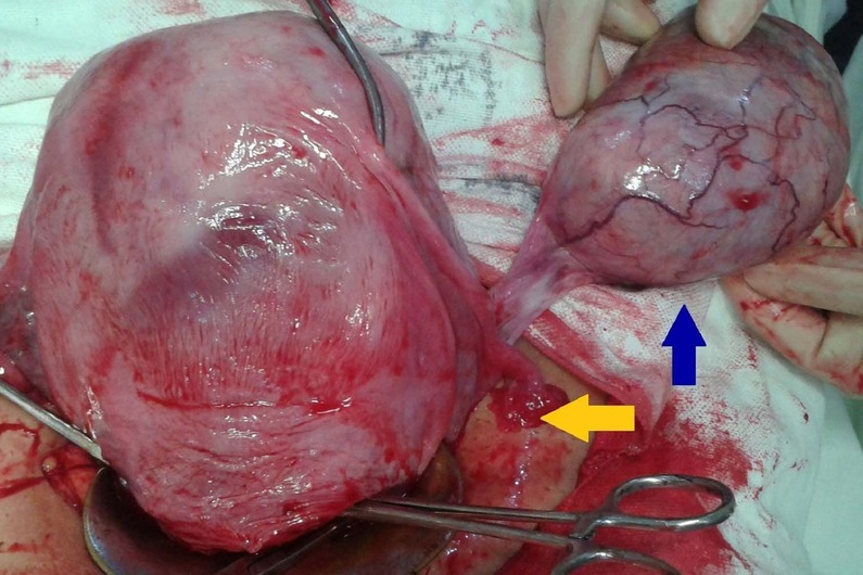
Intraoperative photograph of gravid uterus (yellow arrow) alongside the ovarian tumour (blue arrow) having glistening intact capsule and tortuous vessels on the surface.
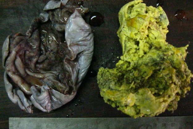
Gross picture showing opened up thin-walled unilocular cyst (left) and its content—yellowish sebaceous material and tufts of hairs (right).

H&E stained sections from the tumour showing (A) Cyst wall lined by keratinised stratified squamous epithelium with hair follicles, sebaceous glands, adipose tissue and smooth muscle bundles (×40). (B) Melanin loaded elongated cells resembling retinal pigment epithelium merging with glial tissue and blood vessels (×40). (C) Higher magnification of the retinal anlage and pigment epithelium (×100). (D) Neuroglial tissue with cell bodies of neurons (×100).
References
-
- Nogales F, Talerman A, Kubik-Huch RA, et al. Germ cell tumours. In: Tavassoli FA, Deville P. World Health Organization classification of tumours. Pathology and genetics of tumours of the breast and female genital organs. Lyon: IARC Press, 2003:168–71
-
- Padubidri VG, Daftary SN. Disorders of the ovary and benign tumours. In: Padubidri VG, Daftary SN, eds. Shaw's textbook of gynaecology. 14th edn New Delhi: Elsevier, 2008:336–48
-
- Dahl N, Gustavson KH, Rune C, et al. Benign ovarian teratomas: an analysis of their cellular origin. Cancer Genet Cytogenet 1990;46:115–23 - PubMed
-
- Ayhan A, Bukulmez O, Genc C, et al. Mature cystic teratomas of the ovary: case series from one institution over 34 years. Eur J Obstet Gynecol Reprod Biol 2000;88:153–7 - PubMed
Publication types
MeSH terms
LinkOut - more resources
Full Text Sources
Other Literature Sources
Medical