. 2014 Apr;196(7):1325-34.
doi: 10.1128/JB.01290-13. Epub 2014 Jan 17.

Expression and targeting of secreted proteins from Chlamydia trachomatis

Affiliations

Expression and targeting of secreted proteins from Chlamydia trachomatis

Laura D Bauler et al. J Bacteriol. 2014 Apr.

Abstract

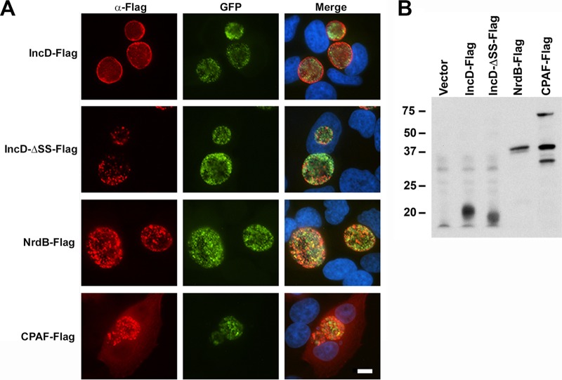
Chlamydia trachomatis is an obligate intracellular pathogen that replicates in a vacuole termed the inclusion. Many of the interactions of chlamydiae with the host cell are dependent upon bacterial protein synthesis and presumably exposure of these proteins to the cytosol. Because of the dearth of genetic tools for chlamydiae, previous studies examining secreted proteins required the use of heterologous bacterial systems. Recent advances in genetic manipulation of chlamydia now allow for transformation of the bacteria with plasmids. We describe here a shuttle vector system, pBOMB4, that permits expression of recombinant proteins under constitutive or conditional promoter control. We show that the inclusion membrane protein IncD is secreted in a type III-dependent manner from Yersinia pseudotuberculosis and also secreted from C. trachomatis in infected cells where it localizes appropriately to the inclusion membrane. IncD truncated of the first 30 amino acids containing the secretion signal is no longer secreted and is retained by the bacteria. Cytosolic exposure of secreted proteins can be confirmed by using CyaA, GSK, or microinjection assays. A protein predicted to be retained within the bacteria, NrdB is indeed localized to the chlamydia. In addition, we have shown that the chlamydial effector protein, CPAF, which is secreted into the host cell cytosol by a Sec-dependent pathway, also accesses the cytosol when expressed from this system. These assays should prove useful to assess the secretion of other chlamydial proteins that are potentially exposed to the cytosol of the host cell.


Figures

FIG 1
FIG 1
pBOMB4 cloning vector for C. trachomatis. Maps of the pBOMB4 vectors. Open reading frames from the C. trachomatis L2 434/Bu vector are labeled pgp1-pgp8. A multiple cloning site containing BamHI, SacII, NotI, NheI, PstI, AgeI, KpnI, and SalI sites was constructed and inserted between the L2 plasmid and β-lactamase gene. The β-lactamase gene, pUC19 origin of replication, and GFP gene were amplified from pGFP-SW2. The pBOMB4R vector is identical to pBOMB4 except that it contains the rpoB promoter (indicated in black). Both pBOMB4 and pBOMB4R have mCherry versions constructed by replacing the GFP gene with an mCherry gene (indicated by gray arrows). The pBOMB4-Tet-mCherry vector contains a tetracycline-inducible promoter and mCherry gene inserted into the BamHI/NotI and NotI/PstI restriction sites of pBOMB4, respectively (indicated by black arrows).
FIG 2
FIG 2
Characterization of C. trachomatis L2 transformed with the pBOMB4 vector. (A) HeLa cells were infected with C. trachomatis L2 (●) or one of the following transformed strains: pBOMB4R-IncD-Flag (○), pBOMB4R-IncD-CyaA (▲), or pBOMB4R (■). At various time points postinfection, cells were lysed in water and replated on fresh monolayers to enumerate the IFU (means ± the standard errors of the mean [SEM]; n = 3). (B) Density gradient-purified EBs were lysed in 20 mM DTT. Quantitative PCR was performed using primers specific to gDNA, the L2 plasmid, or the pBOMB4 plasmid. Copy number was determined relative to genomic DNA. Shown are the means ± the SEM (n = 3).
FIG 3
FIG 3
Tetracycline-inducible promoter driven mCherry expression in C. trachomatis. (A) HeLa cells were infected with C. trachomatis L2 transformed with pBOMB4-Tet-mCherry for 8 h, at which time various amounts of anhydrotetracycline hydrochloride (aTC) were added to induce the expression of mCherry. All mCherry (red) images were taken at the same exposure. Green images indicate the expression of the GFP gene on the plasmid. (B) HeLa cells were infected as described above. At 24 h postinfection, 50 ng of aTC/ml was added to examine the induction of mCherry over time. Bar, 10 μm.
FIG 4
FIG 4
IncD is secreted in a type III secretion system-dependent manner in Yersinia pseudotuberculosis. Y. pseudotuberculosis YPIII pIB102 (WT) or YPIII pIB68 (yscS) expressing IncD, IncD-ΔSS, IncC, NrdB, or vector-only controls were grown in brain heart infusion broth in the presence (+) or absence (−) of 5 mM Ca2+. Cultures were incubated at 26°C, after 2 h protein was induced with 0.01 mM IPTG, and cultures were shifted to 37°C for an additional 4 h. Cultures were fractionated into pelleted cells (P) or supernatant (S or Sup). Samples were resolved on an SDS-PAGE gel and immunoblotted with α-Flag M2 (A) or α-YopN (B).
FIG 5
FIG 5
Secretion of recombinant products from C. trachomatis. HeLa cell monolayers were infected with C. trachomatis L2 transformed with pBOMB4-Tet-IncD-Flag, pBOMB4-Tet-IncD-ΔSS-Flag, pBOMB4-Tet-NrdB-Flag, or pBOMB4-Tet-CPAF-Flag for 24 h before fixation with 4% paraformaldehyde for immunofluorescence (A) or lysed in 2× SDS-PAGE buffer for immunoblotting (B). (A) For immunofluorescence, samples were probed with anti-IncD or α-Flag antibody, followed by a secondary antibody of Alexa 594 (red) and counterstained for DNA with DAPI. Bar, 10 μm. (B) The immunoblot was probed with anti-Flag antibody, followed by an HRP-conjugated secondary antibody. The calculated molecular masses of the flag-tagged fusion proteins were as follows: IncD (19.7 kDa), IncD-ΔSS (16.3 kDa), NrdB (43.2 kDa), and CPAF (70.7 kDa). Two apparent proteolytic fragments of CPAF are also visible.
FIG 6
FIG 6
Cytosolic exposure of proteins secreted by C. trachomatis determined by CyaA. HeLa cells were infected with C. trachomatis L2 transformed with pBOMB4-Tet-IncD-CyaA, pBOMB4-Tet-IncD-ΔSS-CyaA, or pBOMB4-Tet-NrdB-CyaA for 24 h before fixation with 4% paraformaldehyde for immunofluorescence (A) or lysed in 2× SDS-PAGE buffer for immunoblotting (B). Samples were probed with anti-IncD or anti-CyaA antibody, followed by a secondary antibody of Alexa 594 (red) and DAPI (blue) (A) or HRP (B). The approximate sizes for CyaA-tagged proteins are as follows: IncD (60.7 kDa), IncD-ΔSS (57.3 kDa), NrdB (84.2 kDa), and CPAF (111.7 kDa). For the cAMP assay, HeLa cells were infected for 24 h in the presence of 500 µM IBMX and 100 µM Ro 20-1724, at which time the cAMP assay was performed. Fold induction of cAMP was calculated relative to uninfected cells. t test analysis indicates the pBOMB4-Tet-IncD-CyaA cAMP induction is significantly higher than uninfected, IncD-ΔSS and NrdB cAMP levels (P < 0.001; n = 3). Bar, 10 μm.
FIG 7
FIG 7
GSK assay indicates C. trachomatis proteins can be secreted and gain access to the cytosol. HeLa cells were infected with C. trachomatis L2 transformed with pBOMB4-Tet-IncD-GSK, pBOMB4-Tet-IncD-Δ SS-GSK, or pBOMB4-Tet-NrdB-GSK for 24 h before fixation with 4% paraformaldehyde for immunofluorescence (A) or lysed in 2× SDS-PAGE buffer for immunoblotting (B). (A) Samples were probed with α-GSK or α-Phospho-GSK antibody, followed by a secondary antibody of Alexa 594 (red) and DAPI (blue). The α-phospho-GSK antibody shows some cross-reactivity with the endogenous phospho-GSK in the nucleus. Bar, 10 μm. (B) Immunoblots were probed with α-GSK tag or α-phospho-GSK antibody followed by an HRP-conjugated secondary antibody. Approximate sizes for the GSK-IncD, GSK-IncD-Δ SS-GSK, and GSK-NrdB are 18.7, 15.3, and 42.2 kDa, respectively.
FIG 8
FIG 8
Microinjection confirms cytosolic exposure of the IncD-Flag protein. Monolayers of HeLa cells were infected with C. trachomatis L2 transformed with pBOMB4R-IncD-Flag, or pBOMB4R for 3 h before the anti-Flag antibody was microinjected into cells. The cultures were incubated for an additional 21 h before fixation with 4% paraformaldehyde for immunofluorescence. Samples were probed with a secondary antibody of Alexa 594 (red), GFP is expressed from the plasmid in transformed chlamydia (green), and Nomarski (differential interference contrast) images are also shown. Bar, 10 μm.

Similar articles

Cited by

References

    1. Burton MJ, Mabey DC. 2009. The global burden of trachoma: a review. PLoS Negl. Trop. Dis. 3:e460. 10.1371/journal.pntd.0000460 - DOI - PMC - PubMed
    1. Bébéar C, de Barbeyrac B. 2009. Genital Chlamydia trachomatis infections. Clin. Microbiol. Infect. 15:4–10. 10.1111/j.1469-0691.2008.02647.x - DOI - PubMed
    1. Schachter J. 1999. Infection and disease epidemiology, p 139–169 In Stephens RS. (ed), Chlamydia: intracellular biology, pathogenesis, and immunity. ASM Press, Washington, DC
    1. Grayston JT. 1992. Infections caused by Chlamydia pneumoniae strain TWAR. Clin. Infect. Dis. 15:757–761. 10.1093/clind/15.5.757 - DOI - PubMed
    1. Beeckman DS, Vanrompay DC. 2009. Zoonotic Chlamydophila psittaci infections from a clinical perspective. Clin. Microbiol. Infect. 15:11–17. 10.1111/j.1469-0691.2008.02669.x, 10.1111/j.1469-0691.2009.03054.x - DOI - DOI - PubMed

Publication types

MeSH terms

Substances

LinkOut - more resources