

Low-grade myxofibrosarcoma following a metal implantation in femur: a case report
- PMID: 24444015
- PMCID: PMC3926977
- DOI: 10.1186/1746-1596-9-6
Low-grade myxofibrosarcoma following a metal implantation in femur: a case report
Abstract
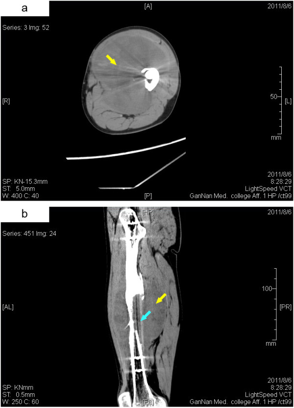
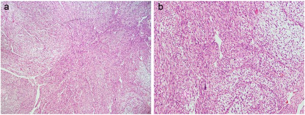
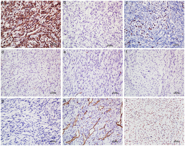
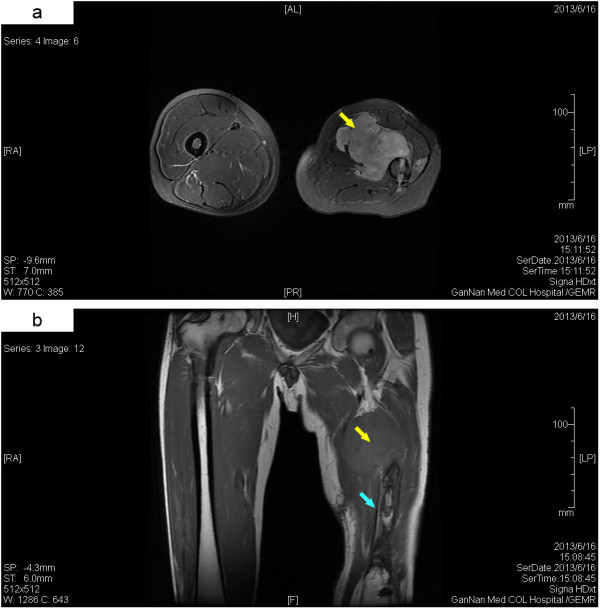
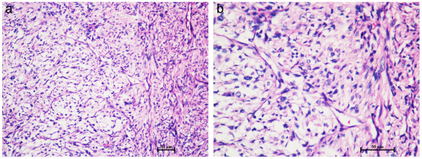
Myxofibrosarcoma is a myxoid variant of malignant fibrous histiocytoma that most commonly involves the extremities of elderly people. However, a primary myxofibrosarcoma with bone invasion in young adults is extremely rare. Herein, we report the case of a 31-year-old male with a gradually enlarging left thigh mass, who had a history of left femur fracture and received an open reduction and internal fixation with titanium alloy plates and screws 33 months previously. Imaging investigations revealed an irregularly shaped soft tissue mass around the left femur shaft and a partial bone defect in the middle one-third of the left femur. Pathological examination of the resected specimen showed a multi-nodular appearance, abundant myxoid matrix and elongated curvilinear capillaries. Immunohistochemical studies revealed that the tumor cells was positive for VIM and MDM2, and was negative for CK, MSA, SMA, DES, S-100 and CD34. Labeling index of Ki-67 was 25%. Based on the morphological finding and immunostaining, it was diagnosed as a low-grade myxofibrosarcoma. The clinical and imaging examinations did not reveal the evidence of a primary cancer elsewhere, and the patient had no personal or family history of malignancy. To our knowledge, this is the first case of a primary myxofibrosarcoma developed following a fracture and metal implantation in young adults.
Virtual slides: The virtual slide(s) for this article can be found here: http://www.diagnosticpathology.diagnomx.eu/vs/1745984882113605.
Figures
Similar articles
-
Diagnosis of extraskeletal myxoid chondrosarcoma in the thigh using EWSR1-NR4A3 gene fusion: a case report.J Med Case Rep. 2016 Nov 10;10(1):321. doi: 10.1186/s13256-016-1113-2. J Med Case Rep. 2016. PMID: 27832806 Free PMC article.
-
CASE REPORTS: malignant fibrous histiocytoma of bone arising in chronic osteomyelitis.Clin Orthop Relat Res. 2005 Oct;439:269-73. doi: 10.1097/01.blo.0000176556.12728.cd. Clin Orthop Relat Res. 2005. PMID: 16205169
-
Myxofibrosarcoma involving brachial plexus diagnoses by contrast-enhanced ultrasound: A case report.Medicine (Baltimore). 2023 Dec 15;102(50):e36626. doi: 10.1097/MD.0000000000036626. Medicine (Baltimore). 2023. PMID: 38115261 Free PMC article.
-
Malignant perivascular epithelioid cell tumor (PEComa) of the femur: a case report and literature review.Diagn Pathol. 2015 May 29;10:54. doi: 10.1186/s13000-015-0292-2. Diagn Pathol. 2015. PMID: 26022435 Free PMC article. Review.
-
Cytopathology of myxofibrosarcoma: a study of 66 cases and literature review.J Am Soc Cytopathol. 2021 May-Jun;10(3):300-309. doi: 10.1016/j.jasc.2020.09.004. Epub 2020 Sep 19. J Am Soc Cytopathol. 2021. PMID: 33041221 Review.
Cited by
-
Surgical technique for reconstruction of diaphyseal defect with endoprosthesis following intercalary resection in femoral shaft.Orthop Surg. 2014 Nov;6(4):329-31. doi: 10.1111/os.12145. Orthop Surg. 2014. PMID: 25430719 Free PMC article. No abstract available.
-
Primary Osseous Low-grade Myxofibrosarcoma of Metatarsal Masquerading as Enchondroma: A Case Report.J Orthop Case Rep. 2022;12(5):35-39. doi: 10.13107/jocr.2022.v12.i05.2806. J Orthop Case Rep. 2022. PMID: 36660161 Free PMC article.
-
Malignant fibrous histiocytoma of the bone in a traumatic amputation stump: A case report and review of the literature.World J Clin Cases. 2021 Sep 16;9(26):7930-7936. doi: 10.12998/wjcc.v9.i26.7930. World J Clin Cases. 2021. PMID: 34621848 Free PMC article.
-
Primary Osseous Low-Grade Myxofibrosarcoma of Clavicle Presenting With Multiple Skeletal Metastases.Cureus. 2020 Aug 31;12(8):e10170. doi: 10.7759/cureus.10170. Cureus. 2020. PMID: 33029450 Free PMC article.
References
-
- Angervall L, Kindblom LG, Merck C. Myxofibrosarcoma. A study of 30 cases. Acta Pathol Microbiol Scand A. 1977;85A(2):127–140. - PubMed
-
- Wang JG, Li YJ, Liu H, Zhao P. Primary cardiac myxofibrosarcoma: a case report and review of the literature. Tumori. 2012;98(6):e165–e168. - PubMed
-
- Kapur P, Sarode V. Pathologic quiz case: myxoid tibial lesion in a 31-year-old man. Low-grade myxofibrosarcoma. Arch Pathol Lab Med. 2004;128(4):e65–e66. - PubMed
Publication types
MeSH terms
Substances
LinkOut - more resources
Full Text Sources
Other Literature Sources
Medical
Research Materials
Miscellaneous
