. 2014 Mar;18(3):396-414.
doi: 10.1111/jcmm.12198. Epub 2014 Jan 20.

Regional mapping of myocardial hibernation phenotype in idiopathic end-stage dilated cardiomyopathy

Affiliations

Regional mapping of myocardial hibernation phenotype in idiopathic end-stage dilated cardiomyopathy

Vincenzo Lionetti et al. J Cell Mol Med. 2014 Mar.

Abstract

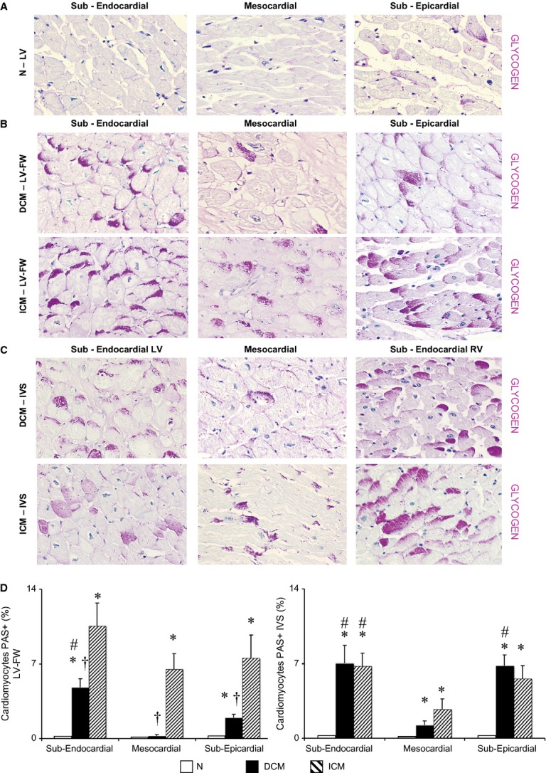
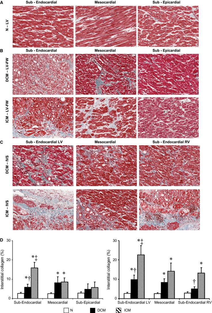
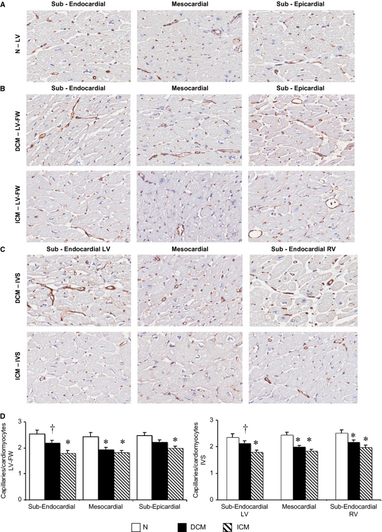
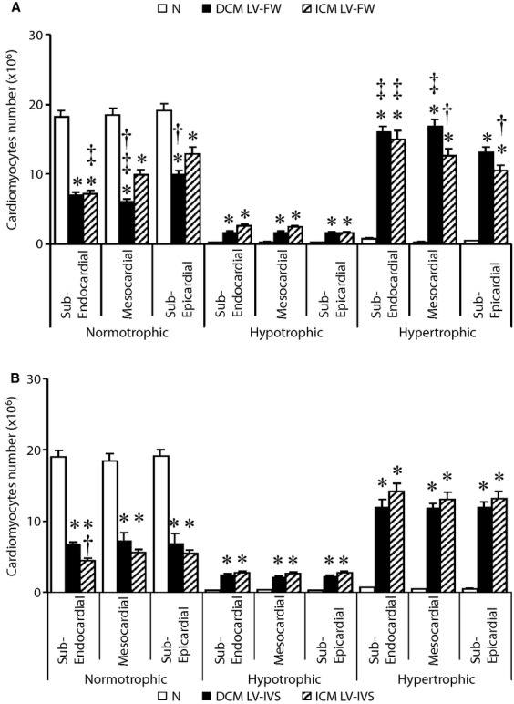
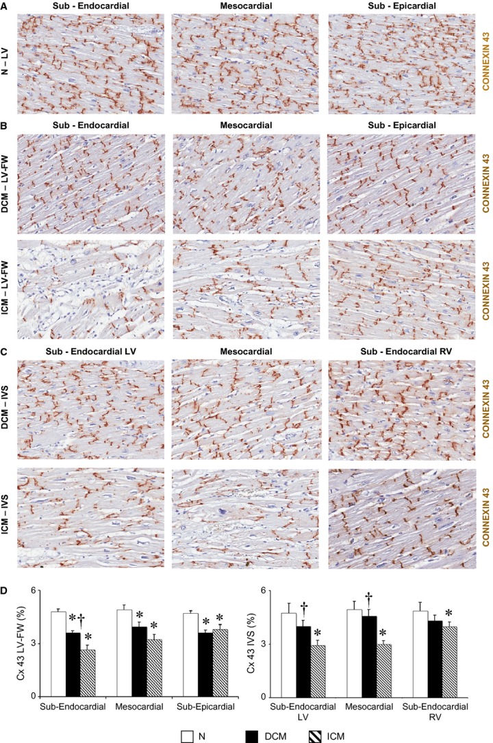
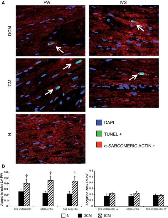
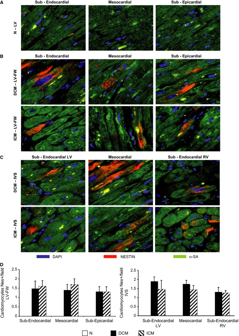
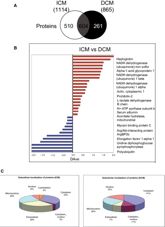
Myocardial hibernation (MH) is a well-known feature of human ischaemic cardiomyopathy (ICM), whereas its presence in human idiopathic dilated cardiomyopathy (DCM) is still controversial. We investigated the histological and molecular features of MH in left ventricle (LV) regions of failing DCM or ICM hearts. We examined failing hearts from DCM (n = 11; 41.9 ± 5.45 years; left ventricle-ejection fraction (LV-EF), 18 ± 3.16%) and ICM patients (n = 12; 58.08 ± 1.7 years; LVEF, 21.5 ± 6.08%) undergoing cardiac transplantation, and normal donor hearts (N, n = 8). LV inter-ventricular septum (IVS) and antero-lateral free wall (FW) were transmurally (i.e. sub-epicardial, mesocardial and sub-endocardial layers) analysed. LV glycogen content was shown to be increased in both DCM and ICM as compared with N hearts (P < 0.001), with a U-shaped transmural distribution (lower values in mesocardium). Capillary density was homogenously reduced in both DCM and ICM as compared with N (P < 0.05 versus N), with a lower decrease independent of the extent of fibrosis in sub-endocardial and sub-epicardial layers of DCM as compared with ICM. HIF1-α and nestin, recognized ischaemic molecular hallmarks, were similarly expressed in DCM-LV and ICM-LV myocardium. The proteomic profile was overlapping by ~50% in DCM and ICM groups. Morphological and molecular features of MH were detected in end-stage ICM as well as in end-stage DCM LV, despite epicardial coronary artery patency and lower fibrosis in DCM hearts. Unravelling the presence of MH in the absence of coronary stenosis may be helpful to design a novel approach in the clinical management of DCM.

Keywords: chronic heart failure; hibernating myocardium; idiopathic dilated cardiomyopathy; ischaemic microenvironment; nestin; pathologic features.


Figures

Figure 1
Figure 1
Extent and distribution of regional left ventricle (LV) glycogen deposits as shown by periodic acid-Schiff (PAS) staining on histological sections. (A–C) Representative images of PAS stained sections for each myocardial layer in the LV-free wall (FW) of normal donor (N) hearts, in the LV-FW and in the inter-ventricular septum (IVS) of either dilated cardiomyopathy (DCM) and ischemic cardiomyopathy (ICM) hearts (magnification 400 × ); (D) Intracellular glycogen amount in LV-FW and IVS of N (n = 8), of DCM (n = 11) and ICM (n = 12) hearts. Values are means ± SEM. *P < 0.05 versus Normal, # versus mesocardial layer, P < 0.05 versus corresponding layer of ICM heart.
Figure 2
Figure 2
Extent and distribution of regional left ventricle (LV) interstitial collagen. (A–C) Representative images of histological Masson's trichrome-stained sections for each myocardial layer in the LV-free wall (FW) of normal donor (N) hearts, in the LV-FW and in the inter-ventricular septum (IVS) of either dilated cardiomyopathy (DCM) or ischaemic cardiomyopathy (ICM) hearts; (D) Interstitial collagen amount in LV-FW and IVS of N (n = 8), of DCM (n = 11) and ICM (n = 12) hearts. Values are means ± SEM. *P < 0.05 versus Normal, P < 0.05 versus corresponding layer of ICM heart, P < 0.05 versus sub-epicardial/sub-endocardial right ventricle (RV) layer.
Figure 3
Figure 3
Regional left ventricle (LV) distribution of coronary capillaries surrounding cardiomyocytes. (A–C) Representative images of coronary capillaries detected with an antibody directed against human von Willebrand Factor (vWF) in sections of normal left ventricle (N, n = 8) and in LVFW and inter-ventricular septum (IVS) of dilated cardiomyopathy (DCM;n = 11) and ischaemic cardiomyopathy (ICM;n = 12) hearts; (D) quantification of the number of capillaries per number of cardiomyocytes per field for each myocardial layer. Values are means ± SEM. *P < 0.05 versus Normal, P < 0.05 versus corresponding layer of ICM heart.
Figure 4
Figure 4
Regional quantification of normo-, hypo-and hyper-trophic cardiomyocytes in each myocardial layer of left ventricle-free wall (LV-FW; A) and inter-ventricular septum (IVS; B) of N (n = 8), dilated cardiomyopathy (DCM;n = 11) and ischaemic cardiomyopathy (ICM;n = 12) hearts. Values are means ± SEM. *P < 0.05 versus Normal, P < 0.05 versus corresponding layer of ICM heart, P < 0.05 versus sub-epicardial layer/sub-endocardial right ventricle (RV) layer.
Figure 5
Figure 5
Extent and distribution of regional left ventricle (LV) connexin-43 (Cx 43). (A–C) Representative images of Cx 43 detected by immunohistochemistry in N LV (n = 8), and in the LVFW and inter-ventricular septum (IVS) of dilated cardiomyopathy (DCM;n = 11) and ischaemic cardiomyopathy (ICM;n = 12) hearts; (D) quantification of immunodetectable Cx 43 in each LV myocardial layer. Values are expressed as means ± SEM. *P < 0.05 versus Normal, P < 0.05 versus corresponding layer of ICM heart.
Figure 6
Figure 6
Regional detection of ventricular apoptotic cardiomyocytes (α-SA-positive cells) in left ventricle-free wall (LV-FW) and in inter-ventricular septum (IVS) of dilated cardiomyopathy (DCM;n = 11) and ischaemic cardiomyopathy (ICM;n = 11) hearts, and N hearts (n = 8) by TUNEL assay (A) and regional LV apoptotic index (B). α-SA: alpha-sarcomeric actinin. Values are expressed as means ± SEM. P < 0.05 versus corresponding LV layer of DCM heart.
Figure 7
Figure 7
Regional detection of ventricular nestin-positive cardiomyocytes. (A–C) Representative immunofluorescence sections of nestin-positive cardiomyocytes (α-SA-positive cells) in each left ventricle (LV) myocardial layer of N (n = 8) and LVFW and inter-ventricular septum (IVS) of dilated cardiomyopathy (DCM;n = 11) and ischaemic cardiomyopathy (ICM;n = 12) hearts; (D) quantification of nestin-positive cardiomyocytes in each LV myocardial layer of both heart failure groups. α-SA: alpha-sarcomeric actinin. Values are expressed as means ± SEM.
Figure 8
Figure 8
Proteomic profile. (A) Schematic representation of human proteome in inter-ventricular septum (IVS) of both (ischaemic cardiomyopathy, ICM and dilated cardiomyopathy, DCM) failing heart groups showing homology of myocardial proteins. (B) Semiquantitative analysis, in which the bars represent the score ratio expressed by the DAve index. (C) Schematic representation of subcellular distribution of myocardial proteins. n = 5 hearts per ICM and DCM group.

References

    1. Canty JM, Jr, Suzuki G. Myocardial perfusion and contraction in acute ischemia and chronic ischemic heart disease. J Mol Cell Cardiol. 2012;52:822–31. - PMC - PubMed
    1. Heusch G. Hibernating myocardium. Physiol Rev. 1998;78:1055–85. - PubMed
    1. Skalidis EI, Parthenakis FI, Patrianakos AP, et al. Regional coronary flow and contractile reserve in patients with idiopathic dilated cardiomyopathy. J Am Coll Cardiol. 2004;44:2027–32. - PubMed
    1. Baumgartner H, Porenta G, Lau YK, et al. Assessment of myocardial viability by dobutamine echocardiography, positron emission tomography and thallium-201 SPECT: correlation with histopathology in explanted hearts. J Am Coll Cardiol. 1998;32:1701–8. - PubMed
    1. Elsasser A, Vogt AM, Nef H, et al. Human hibernating myocardium is jeopardized by apoptotic and autophagic cell death. J Am Coll Cardiol. 2004;43:2191–9. - PubMed

Publication types

MeSH terms

Supplementary concepts

LinkOut - more resources