. 2014 Mar;59(100):24-36.
doi: 10.1016/j.mcn.2014.01.002. Epub 2014 Jan 18.

Selective regulation of axonal growth from developing hippocampal neurons by tumor necrosis factor superfamily member APRIL

Affiliations

Selective regulation of axonal growth from developing hippocampal neurons by tumor necrosis factor superfamily member APRIL

Catarina Osório et al. Mol Cell Neurosci. 2014 Mar.

Abstract

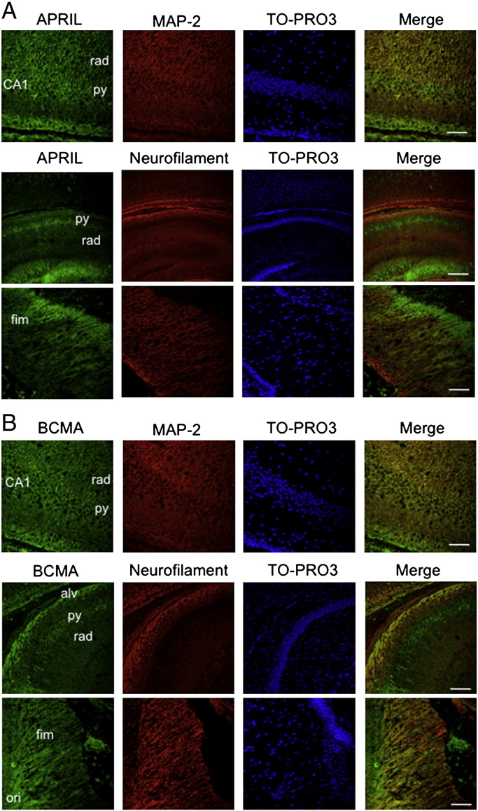
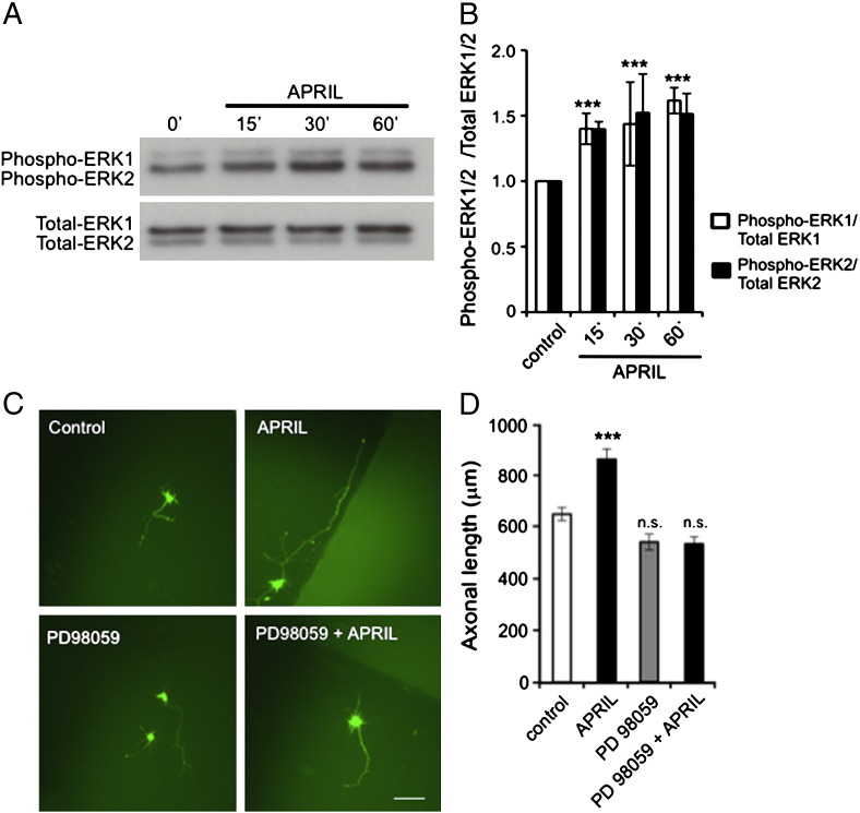
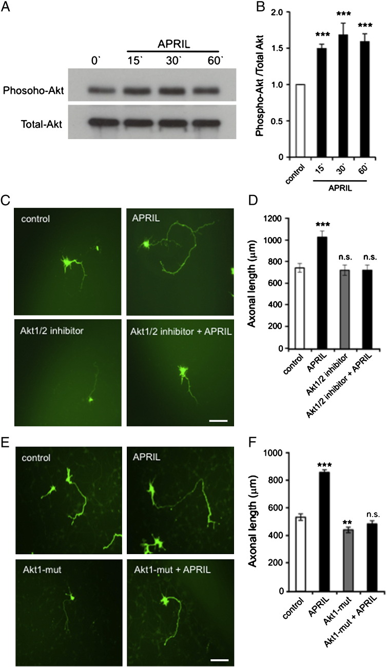
APRIL (A Proliferation-Inducing Ligand, TNFSF13) is a member of the tumor necrosis factor superfamily that regulates lymphocyte survival and activation and has been implicated in tumorigenesis and autoimmune diseases. Here we report the expression and first known activity of APRIL in the nervous system. APRIL and one of its receptors, BCMA (B-Cell Maturation Antigen, TNFRSF17), are expressed by hippocampal pyramidal cells of fetal and postnatal mice. In culture, these neurons secreted APRIL, and function-blocking antibodies to either APRIL or BCMA reduced axonal elongation. Recombinant APRIL enhanced axonal elongation, but did not influence dendrite elongation. The effect of APRIL on axon elongation was inhibited by anti-BCMA and the expression of a signaling-defective BCMA mutant in these neurons, suggesting that the axon growth-promoting effect of APRIL is mediated by BCMA. APRIL promoted phosphorylation and activation of ERK1, ERK2 and Akt and serine phosphorylation and inactivation of GSK-3β in cultured hippocampal pyramidal cells. Inhibition of MEK1/MEK2 (activators of ERK1/ERK2), PI3-kinase (activator of Akt) or Akt inhibited the axon growth-promoting action of APRIL, as did pharmacological activation of GSK-3β and the expression of a constitutively active form of GSK-3β. These findings suggest that APRIL promotes axon elongation by a mechanism that depends both on ERK signaling and PI3-kinase/Akt/GSK-3β signaling.

Keywords: Axon; Hippocampal pyramidal cell; Tumor necrosis factor receptor superfamily; Tumor necrosis factor superfamily.


Figures

Fig. 1
Fig. 1
Expression of APRIL and BCMA in the developing hippocampus. Graphs of the levels of APRIL mRNA (A) and BCMA mRNA (B) relative to reference mRNAs for GAPDH and SDHA in total RNA extracted from E18, P0, P5 and P10 hippocampi (mean ± s.e.m., n = 4 separate sets of hippocampal tissue at each age). Representative Western blots of lysates of E18, P0, P5, and P10 hippocampi probed for GAPDH together with either APRIL (C) or BCMA (D). Lysates from spleen tissue were probed as positive control.
Fig. 2
Fig. 2
Localization of APRIL and BCMA in the developing hippocampus. Frozen sections of P10 hippocampus triple labeled with TO-PRO®-3 Iodide, anti-MAP-2 or anti-200 kD neurofilament and either (A) anti-APRIL (B) or anti-BCMA. For each set, the upper panels show a higher power image of CA1 (scale bars = 25 μm), the middle panels show lower power images of the CA regions (scale bars = 200 μm) and the lower panel show a higher power image of the fimbria (scale bars = 25 μm). Abbreviations: rad, stratum radiatum; py, stratum pyramidale; fim, fimbria; alv, alveus; ori, stratum oriens.
Fig. 3
Fig. 3
APRIL selectively enhances axonal growth from cultured hippocampal pyramidal neurons. (A–C) Axonal growth from E18 hippocampal pyramidal neurons after 3 days in vitro. The neurons were transfected 2 days after plating with a plasmid expressing GFP and were treated with APRIL for the last 18 h of culture prior to fixation and immunostaining for GFP. (A) Representative neurons incubated without APRIL (Control) or treated with 100 ng/ml APRIL. Scale bar = 100 μm. (B) Axon length in control cultures and cultures treated with APRIL at the concentrations indicated. (C) Camera lucida drawings of control neurons and neurons treated with 100 ng/ml APRIL. The neurons illustrated correspond to the 100th, 75th, 50th and 25th percentiles in terms of total neurite length for each condition. (D) Representative E18 CA1 explants incubated with and without APRIL (100 ng/ml) for 3 days before labeling the emergent neurite with the fluorescent vital dye calcein AM. Scale bar = 200 μm. (E–F) Dendrite growth from E18 hippocampal pyramidal neurons after 7 days in vitro. The neurons were transfected with pGFP 6 days after plating and were incubated with and without APRIL for the last 18 h of culture prior to fixation and immunostaining for GFP. (E) Representative neurons incubated without APRIL (Control) or treated with 100 ng/ml APRIL. Scale bar = 50 μm. (F) Bar chart of the percentage of dendrites longer than 50 μm in control cultures and cultures treated with APRIL at the concentrations indicated. The data shown in B and F represent the mean ± s.e.m. of data compiled from > 150 neurons per condition from at least three separate experiments (*** indicates P < 0.0001, statistical comparison with control, Mann–Whitney U test).
Fig. 4
Fig. 4
BCMA mediates APRIL-promoted axonal outgrowth. (A) Representative E18 hippocampal pyramidal neurons that were transfected after 2 days in vitro with plasmids expressing either GFP alone or GFP together with either wild-type BCMA or truncated BCMA (BCMA ΔC86). After transfection, the neurons were cultured for a further 18 h with and without APRIL (100 ng/ml) before GFP immunostaining. Scale bar = 100 μm. (B) Bar chart of axonal length under the experimental conditions illustrated in A. (C) Bar chart of axonal length of neurons cultured in the presence or absence of 3 μg/ml of anti-BCMA or 1.5 μg/ml of anti-APRIL with or without APRIL (100 ng/ml). The mean ± s.e.m. of data obtained from > 150 neurons per condition compiled from at least three separate experiments are shown (*** indicates P < 0.0001, statistical comparison with control, Mann–Whitney U test).
Fig. 5
Fig. 5
APRIL promotes axonal growth by activating ERK1 and ERK2. (A) Representative Western blots probed for phospho-ERK1/ERK2 and total ERK1/ERK2 of lysates of E18 hippocampal neurons of treated with 100 ng/ml APRIL for the times indicated after first culturing the neurons for 3 days. (B) Densitometric quantification of levels of phospho-ERK1/phospho-ERK2 relative to total ERK1/total ERK2 from 3 separate Western blot experiments (mean ± s.e.m., *** indicates P < 0.0001, statistical comparison with control, Mann–Whitney U test). (C) Images of representative E18 hippocampal pyramidal neurons that were transfected after 2 days in vitro with a plasmid expressing GFP and were treated with 10 μM of PD98059 2 h prior to incubation for 18 h with and without APRIL (100 ng/ml) before GFP immunostaining. Separate cultures were treated with APRIL after transfection and control cultures received neither PD98059 nor APRIL. Scale bar = 100 μm. (D) Bar chart of the axonal growth under the experimental conditions illustrated in C. The mean ± s.e.m. of data obtained from > 150 neurons per condition compiled from at least three separate experiments are shown (*** indicates P < 0.0001 and n.s., non-significant, statistical comparison with control, Mann–Whitney U test).
Fig. 6
Fig. 6
APRIL-promoted axonal growth depends on PI-3 kinase activation. (A) Images of representative E18 hippocampal pyramidal neurons that were transfected after 2 days in vitro with a plasmid expressing GFP and were treated with 10 μM of LY294002 2 h prior to a further 18 h of incubation with and without APRIL (100 ng/ml) before GFP immunostaining. Separate cultures were treated with APRIL after transfection and control cultures received neither LY294002 nor APRIL. (B) Bar chart of the axonal growth under the experimental conditions illustrated in A. (C) Images of representative E18 hippocampal pyramidal neurons that were transfected after 2 days in vitro with plasmids expressing either GFP alone or GFP together with a dominant-negative form of p85α regulatory subunit of PI-3 kinase (p85α-DN). After transfection, the neurons were cultured for a further 18 h with and without APRIL (100 ng/ml) before GFP immunostaining. (D) Bar chart of the axonal growth under the experimental conditions illustrated in C. The mean ± s.e.m. of data obtained from > 150 neurons per condition compiled from at least three separate experiments are shown in B and D (** indicates P < 0.001, *** indicates P < 0.0001 and n.s., non-significant, statistical comparison with control, Mann–Whitney U test). Scale bars = 100 μm.
Fig. 7
Fig. 7
APRIL promotes axonal growth by activating Akt signaling. (A) Representative Western blot probed for phospho-Akt and total Akt in lysates of E18 hippocampal neurons of treated with 100 ng/ml APRIL for the times indicated after first culturing the neurons for 3 days. (B) Densitometric quantification of levels of phospho-Akt relative to total Akt from 3 separate Western blot experiments (mean ± s.e.m., *** indicates P < 0.0001, statistical comparison with control, Mann–Whitney U test). (C) Images of representative E18 hippocampal pyramidal neurons that were transfected after 2 days in vitro with a plasmid expressing GFP and were treated with 5 μM of the Akt1/2 inhibitor 2 h prior to a further 18 h incubation with and without APRIL (100 ng/ml) before GFP immunostaining. Separate cultures were treated with APRIL after transfection and control cultures received neither the inhibitor nor APRIL. (D) Bar chart of the axonal growth under the experimental conditions illustrated in C. (E) Images of representative E18 hippocampal pyramidal neurons that were transfected after 2 days in vitro with plasmids expressing either GFP alone or GFP together with the Akt1 K179M T308A S473A mutant (Akt1-mut). After transfection, the neurons were cultured for a further 18 h with and without APRIL (100 ng/ml) before GFP immunostaining. (F) Bar chart of the axonal growth under the experimental conditions illustrated in E. The data shown in D and F are compiled from > 150 neurons per condition from at least three separate experiments (mean ± s.e.m., *** indicates P < 0.0001 and n.s. non-significant, statistical comparison with control, Mann–Whitney U test). Scale bars = 100 μm.
Fig. 8
Fig. 8
APRIL promotes axonal growth by activating GSK-3β signaling. (A) Representative Western blots probed for phospho-GSK-3α, phospho-GSK-3β and total GSK-3β in lysates of E18 hippocampal neurons treated with 100 ng/ml APRIL for the times indicated after first culturing the neurons for 3 days. (B) Densitometric quantification of levels of phospho-GSK-3β relative to total GSK-3β from 3 separate Western blot experiments (mean ± s.e.m., *** indicates P < 0.0001, statistical comparison with control, Mann–Whitney U test). (C) Images of representative E18 hippocampal pyramidal neurons that were transfected after 2 days in vitro plasmids expressing either GFP alone or GFP together with the GSK3β S9A mutant. After transfection, the neurons were cultured for a further 18 h with and without APRIL (100 ng/ml) before GFP immunostaining. (D) Bar chart of the axonal growth under the experimental conditions illustrated in C. The mean ± s.e.m. of data obtained from > 150 neurons per condition compiled from at least three separate experiments are shown in D (*** indicates P < 0.0001 and n.s., non-significant, statistical comparison with control, Mann–Whitney U test). Scale bars = 100 μm.

Similar articles

Cited by

References

    1. Aggarwal B.B. Signalling pathways of the TNF superfamily: a double-edged sword. Nat. Rev. Immunol. 2003;3:745–756. - PubMed
    1. Alexaki V.I., Notas G., Pelekanou V., Kampa M., Valkanou M., Theodoropoulos P., Stathopoulos E.N., Tsapis A., Castanas E. Adipocytes as immune cells: differential expression of TWEAK, BAFF, and APRIL and their receptors, Fn14, BAFF-R, TACI, and BCMA, at different stages of normal and pathological adipose tissue development. J. Immunol. 2009;183:5948–5956. - PubMed
    1. Atwal J.K., Massie B., Miller F.D., Kaplan D.R. The TrkB-Shc site signals neuronal survival and local axon growth via MEK and P13-kinase. Neuron. 2000;27:265–277. - PubMed
    1. Bossen C., Schneider P. BAFF, APRIL and their receptors: structure, function and signaling. Semin. Immunol. 2006;18:263–275. - PubMed
    1. Boukhaddaoui H., Sieso V., Scamps F., Valmier J. An activity-dependent neurotrophin-3 autocrine loop regulates the phenotype of developing hippocampal pyramidal neurons before target contact. J. Neurosci. 2001;21:8789–8797. - PMC - PubMed

Publication types

MeSH terms

Substances