Review
. 2014 Apr:179:49-56.
doi: 10.1016/j.chemphyslip.2013.12.009. Epub 2014 Jan 17.

The functions of cardiolipin in cellular metabolism-potential modifiers of the Barth syndrome phenotype

Affiliations
Review

The functions of cardiolipin in cellular metabolism-potential modifiers of the Barth syndrome phenotype

Vaishnavi Raja et al. Chem Phys Lipids. 2014 Apr.

Abstract

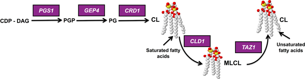
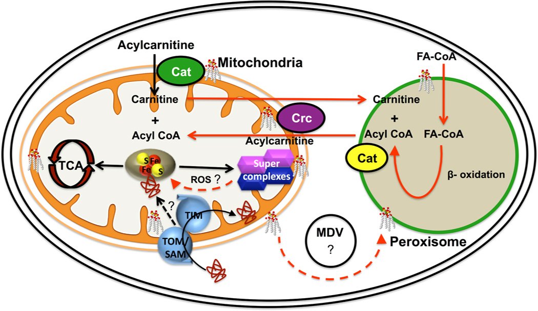
The phospholipid cardiolipin (CL) plays a role in many cellular functions and signaling pathways both inside and outside of mitochondria. This review focuses on the role of CL in energy metabolism. Many reactions of electron transport and oxidative phosphorylation, the transport of metabolites required for these processes, and the stabilization of electron transport chain supercomplexes require CL. Recent studies indicate that CL is required for the synthesis of iron-sulfur (Fe-S) co-factors, which are essential for numerous metabolic pathways. Activation of carnitine shuttle enzymes that are required for fatty acid metabolism is CL dependent. The presence of substantial amounts of CL in the peroxisomal membrane suggests that CL may be required for peroxisomal functions. Understanding the role of CL in energy metabolism may identify physiological modifiers that exacerbate the loss of CL and underlie the variation in symptoms observed in Barth syndrome, a genetic disorder of CL metabolism.

Keywords: Barth syndrome; Bioenergetics; Carnitine transport; Fatty acid utilization; Iron–sulfur biogenesis.


Figures

Figure 1
Figure 1. Synthesis and remodeling of cardiolipin (CL) in yeast
CDP-DAG is converted to phosphatidylglycerolphosphate (PGP) by phosphatidylglycerolphosphate synthase (PGPS), encoded by PGS1 (Chang et al., 1998; Dzugasova et al., 1998). PGP phosphatase (Gep4) catalyzes the conversion of PGP to phosphatidylgylcerol (PG) (Osman et al., 2010). PG is converted to cardiolipin (CL) by CL synthase (Crd1) (Jiang et al., 1997; Chang et al., 1998; Tuller et al., 1998). CL is remodeled in a two-step process in which the CL specific deacylase encoded by CLD1 removes a fatty acyl group, forming monolysocardiolipin (MLCL) (Beranek et al., 2009), and tafazzin (Taz1) reacylates MLCL to form a generally more unsaturated CL (Xu et al., 2003). In mammalian cells, CL is deacylated by more than one enzyme (Kiebish et al., 2013). Tafazzin is the enzyme that is mutated in Barth syndrome.
Figure 2
Figure 2. Functions of cardiolipin (CL) in metabolic pathways
CL is most abundant in the inner membrane and is also present in the outer membrane of mitochondria. It is required for activities of transporters and electron transport chain enzymes and for stabilization of electron transport supercomplexes. Loss of CL leads to perturbation of Fe-S biogenesis, resulting in decreased activity of Fe-S enzymes in the TCA cycle, electron transport, and other pathways. The mechanism linking CL and Fe-S biogenesis is unknown. Because CL is required for the import of proteins through mitochondrial import complexes (TOM, SAM and TIM), it is possible that import of specific proteins required for Fe-S synthesis is defective in CL deficient cells. Alternatively, increased ROS generated by inefficient electron transport in CL deficient cells may damage Fe-S proteins. CL is also present in the membrane of the peroxisome, an organelle that carries out β-oxidation of fatty acids, ether lipid synthesis, and reactions of the glyoxylate cycle. The route whereby CL is transported from mitochondria to peroxisomes is unclear, but may involve mitochondria derived vesicles (MDVs). Acyl CoA produced by β-oxidation of long chain fatty acids in peroxisomes is transported to the mitochondria via the carnitine shuttle. The acyl CoA is transferred to carnitine in the peroxisome by carnitine acyltransferase (Cat). Acylcarnitine from the peroxisome crosses the mitochondrial membrane, facilitated by the carnitine/acylcarnitine translocase (Crc). CL is required for efficient activity of both mitochondrial carnitine enzymes in mammalian cells.

References

    1. Acehan D, Malhotra A, Xu Y, Ren M, Stokes DL, Schlame M. Cardiolipin affects the supramolecular organization of ATP synthase in mitochondria. Biophysical journal. 2011;100:2184–2192. - PMC - PubMed
    1. Balk J, Aguilar Netz DJ, Tepper K, Pierik AJ, Lill R. The essential WD40 protein Cia1 is involved in a late step of cytosolic and nuclear iron-sulfur protein assembly. Molecular and cellular biology. 2005;25:10833–10841. - PMC - PubMed
    1. Balk J, Pierik AJ, Netz DJ, Muhlenhoff U, Lill R. The hydrogenase-like Nar1p is essential for maturation of cytosolic and nuclear iron-sulphur proteins. The EMBO journal. 2004;23:2105–2115. - PMC - PubMed
    1. Barja G. Mitochondrial oxygen radical generation and leak: sites of production in states 4 and 3, organ specificity, and relation to aging and longevity. Journal of bioenergetics and biomembranes. 1999;31:347–366. - PubMed
    1. Bartlett K, Eaton S. Mitochondrial beta-oxidation. European journal of biochemistry / FEBS. 2004;271:462–469. - PubMed

Publication types

LinkOut - more resources