. 2014:5:3164.
doi: 10.1038/ncomms4164.

Maspin is not required for embryonic development or tumour suppression

Affiliations
Free PMC article

Maspin is not required for embryonic development or tumour suppression

Sonia S Y Teoh et al. Nat Commun. 2014.
Free PMC article

Abstract

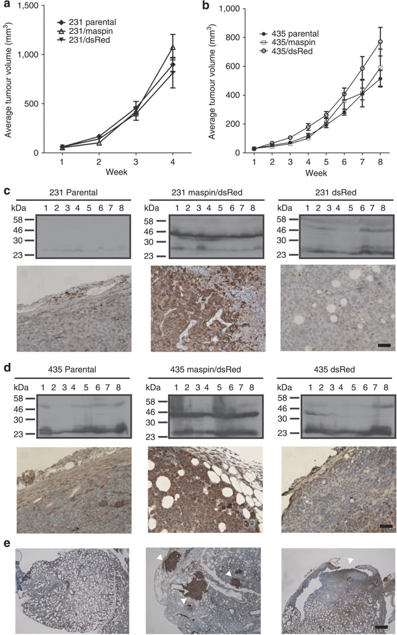
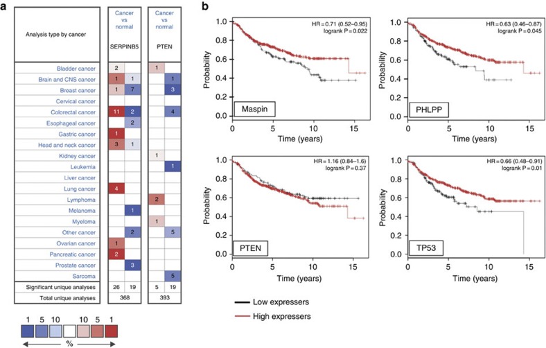
Maspin (SERPINB5) is accepted as an important tumour suppressor lost in many cancers. Consistent with a critical role in development or differentiation maspin knockout mice die during early embryogenesis, yet clinical data conflict on the prognostic utility of maspin expression. Here to reconcile these findings we made conditional knockout mice. Surprisingly, maspin knockout embryos develop into overtly normal animals. Contrary to original reports, maspin re-expression does not inhibit tumour growth or metastasis in vivo, or influence cell migration, invasion or survival in vitro. Bioinformatic analyses reveal that maspin is not commonly under-expressed in cancer, and that perturbation of genes near maspin may in fact explain poor survival in certain patient cohorts with low maspin expression.


Figures

Figure 1
Figure 1. Construction and validation of maspin knockout mice.
(a) Structure of the maspin locus on mouse chromosome 1. (b) Diagrams of the maspin gene and predicted gene structure after insertion of the targeting vector (Serpinb5tm1Pib (targ)); deletion of exon 4 and the neomycin (neo) cassette after exposure to cre recombinase (Serpinb5tm1.1Pib (flox)) or deletion of the neo cassette alone after exposure to flp recombinase (Serpinb5tm1.2Pib (FLP)). Sequences cloned and used as external or internal (int.) probes (pr) are indicated. Coordinates are from the mouse genomic reference sequence GRCm38.p1 C57/BL6. (c) Validation of mouse genotypes by Southern blotting. DNA from wild type (wt) and Serpinb5tm1Pib/Serpinb5wt embryonic stem (ES) cells was used as a control. (d) Structure of maspin mRNA in ko tissue. RT–PCR product from mammary tissue was sequenced directly. Shown is a portion of the sequencing trace covering the deletion break point, with the nucleotide and inferred peptide sequences compared with the reference wt sequence below.
Figure 2
Figure 2. Maspin knockout mice are ostensibly normal.
(a) Comparison of maspin mRNA in adult wild-type (wt) and Cre-deleted knockout (ko) mice. Reverse transcriptase PCR analysis demonstrates products of predicted size (wt: 602 bp; ko: 455 bp), validated by DNA sequencing (see Fig. 1d). (b) ko mice do not produce maspin protein, as assessed by immunoblotting using the anti-human maspin monoclonal antibody 13G11 or rabbit anti-peptide antibody H130. Validation of these antibodies is shown in Supplementary Fig. 1. (c) Maspin ko skin lacks maspin in epidermal cells, as assessed by immunohistochemistry of samples from 8-week old mice, using 13G11. Scale bar, 30 μm.
Figure 3
Figure 3. Maspin does not affect tumour growth, size, morphology or metastasis.
Balb/c nu/nu mice were implanted orthotopically with parental or transduced (maspin/dsRed or dsRed) MDA-MB-231 or MDA-MB-435 cells to generate breast tumour xenografts (six groups, n=8 per group). (a,b) Developing mammary fat pad tumours were measured twice weekly. (c) MDA-MB-231 derived tumours were examined for maspin via immunoblotting (upper panels) and immunohistochemistry (lower panels) using the monoclonal antibody 16F7. Scale bar, 50 μm. (d) MDA-MB-435-derived tumours were examined for maspin via immunoblotting (upper panels) and immunohistochemistry (lower panels) using the monoclonal antibody 16F7. Scale bar, 50 μm. (e) Micrometastatic lesions (arrowheads) were evident in all 435 xenograft types, and these lesions were positive for maspin in the 435/maspin tumours. Scale bar, 200 μm.
Figure 4
Figure 4. Maspin can be over- or under-expressed in cancer.
(a) Differential maspin expression between normal breast tissue and various types of cancers based on default threshold P-value and fold changes set by Oncomine. Over-expression or under-expression in the top 1, 5 and 10% are colour-coded according to the legend. (b) Correlation between lower maspin expression (Affymetrix ID 204855_at (SERPINB5)) and poor recurrence-free survival (RFS) in some breast cancer patients. Plots were generated online using a Kaplan–Meier Plotter based on signal intensity in microarray gene expression data from patients for which recurrence-free survival data is available. Shown are plots for patients in the lower RFS quartile, and comparison to PHLPP (Affymetrix ID 212719_at (PHLPP1)), PTEN (Affymetrix ID 217492_s_at (PTEN)) and TP53 (Affymetrix ID 201746_at (TP53)).

References

    1. Liang P., Averboukh L., Keyomarsi K., Sager R. & Pardee A. B. Differential display and cloning of messenger RNAs from human breast cancer versus mammary epithelial cells. Cancer Res. 52, 6966–6968 (1992). - PubMed
    1. Zou Z. et al.. Maspin, a serpin with tumor-suppressing activity in human mammary epithelial cells. Science 263, 526–529 (1994). - PubMed
    1. Sheng S. et al.. Maspin acts at the cell membrane to inhibit invasion and motility of mammary and prostatic cancer cells. Proc. Natl Acad. Sci. USA 93, 11669–11674 (1996). - PMC - PubMed
    1. Seftor R. E. et al.. Maspin suppresses the invasive phenotype of human breast carcinoma. Cancer Res. 58, 5681–5685 (1998). - PubMed
    1. Cella N., Contreras A., Latha K., Rosen J. M. & Zhang M. Maspin is physically associated with β1 integrin regulating cell adhesion in mammary epithelial cells. FASEB J. 20, 1510–1512 (2006). - PubMed

Publication types

LinkOut - more resources