. 2014 May;133(5):628e-637e.
doi: 10.1097/PRS.0000000000000102.

The effects of biological lubricating molecules on flexor tendon reconstruction in a canine allograft model in vivo

Affiliations

The effects of biological lubricating molecules on flexor tendon reconstruction in a canine allograft model in vivo

Chunfeng Zhao et al. Plast Reconstr Surg. 2014 May.

Abstract

Background: Using allograft is an attractive alternative for flexor tendon reconstruction because of the lack of donor-site morbidity, and better matching to the intrasynovial environment. The purpose of this study was to use biological lubricant molecules to modify the graft surface to decrease adhesions and improve digit function.

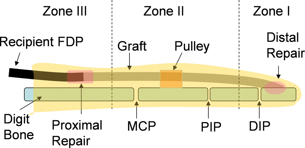
Methods: Twenty-eight flexor digitorum profundus tendons from the second and fifth digits of 14 dogs were lacerated and repaired to create a model with repair failure and scar digit for tendon reconstruction. Six weeks after the initial operation, the tendons were reconstructed with flexor digitorum profundus allograft tendons obtained from canine cadavers. One graft tendon in each dog was treated with saline as a control and the other was treated with carbodiimide-derivatized hyaluronic acid and gelatin plus lubricin. Six weeks postoperatively, digit function, graft mechanics, and biology were analyzed.

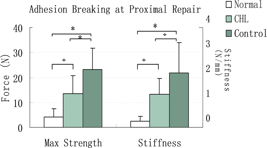
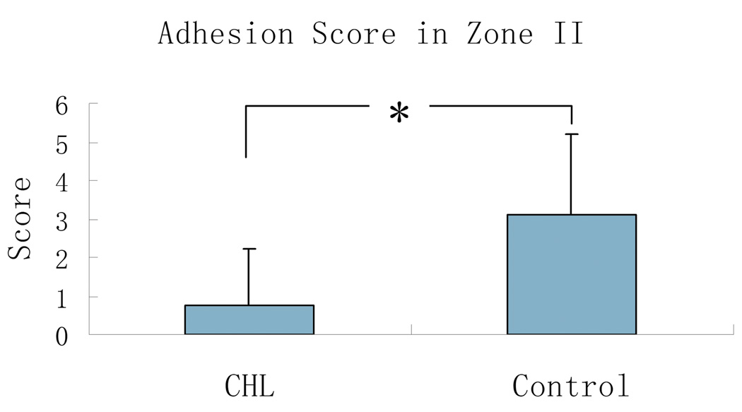
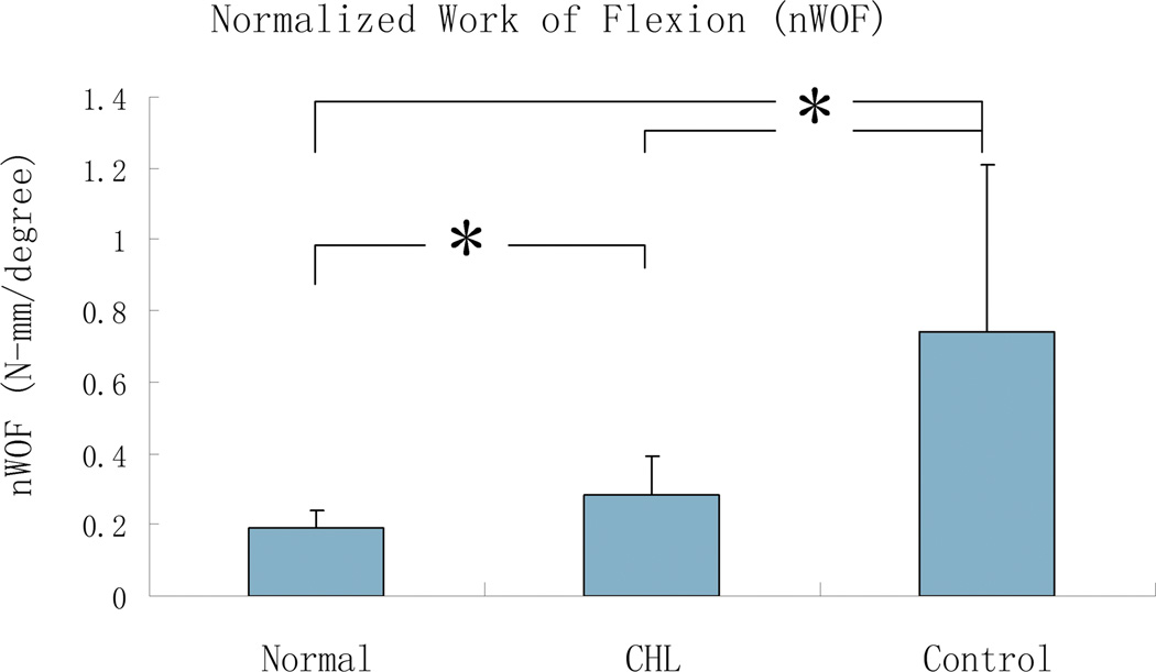
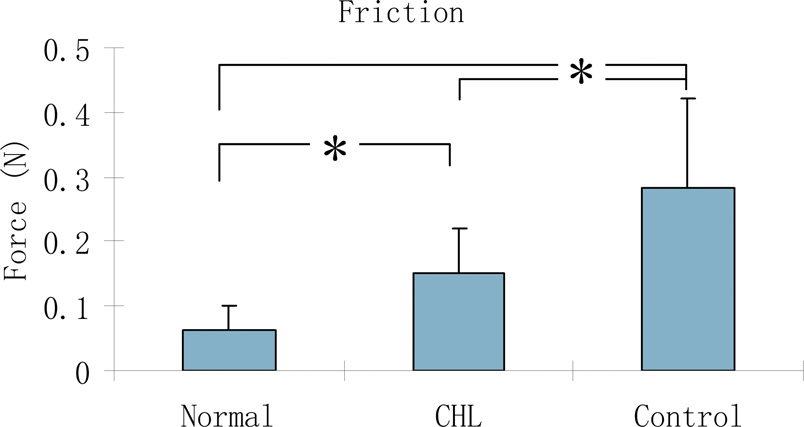
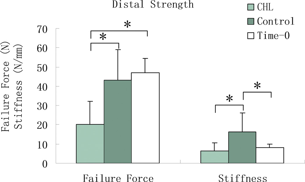
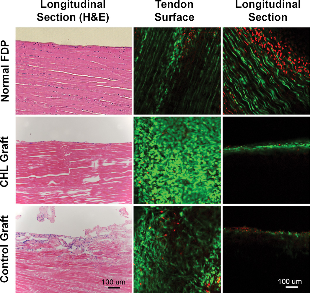
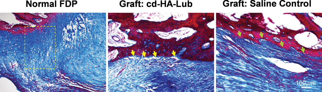
Results: Allograft tendons treated with carbodiimide-derivatized hyaluronic acid-lubricin had decreased adhesions at the proximal tendon/graft repair and within the flexor sheath, improved digit function, and increased graft gliding ability. The treatment also reduced the strength at the distal tendon-to-bone repair, but the distal attachment rupture rate was similar for both graft types. Histologic evaluation showed that viable cells migrated to the allograft, but these were limited to the tendon surface.

Conclusions: Carbodiimide-derivatized hyaluronic acid-lubricin treatment of tendon allograft improves digit functional outcomes after flexor tendon reconstruction. However, delayed bone-to-tendon healing should be a caution. Furthermore, the cell infiltration into the allograft tendon substance should be a target for future studies, to shorten the allograft self-regeneration period.


Figures

Figure 1
Figure 1
Schematic diagram of the graft with digits zones I-III. The graft in zone I and zone II was assessed for work of flexion and adhesion formation. The graft in zone I was also tested for tendon to bone healing strength, and the graft in zone II was assessed for graft friction. The proximal host tendon to graft repair in zone III was used to evaluate the adhesion breaking strength. The graft segments in zone I, II, and III were also processed for histology. (DIP: distal interphalangeal joint; PIP: proximal interphalangeal joint; MCP: metacarpophalangeal joint)
Figure 2
Figure 2
The mean and standard deviation of adhesion breaking strength and stiffness at the proximal tendon/graft repair site for normal FDP tendon (Normal), cd-HA-Lubricin (CHL) treated, and saline treated control graft tendons. Asterisk denotes a significant difference (p < 0.05).
Figure 3
Figure 3
The mean and standard deviation of adhesion score between cd-HA-Lubricin (CHL) and saline control group. Asterisk denotes a significant difference (p < 0.05).
Figure 4
Figure 4
The mean and standard deviation of normalized work of flexion (nWOF) for normal digit (Normal), cd-HA-Lubricin (CHL), and saline treated control groups. Asterisk denotes a significant difference (p < 0.05).
Figure 5
Figure 5
The mean and standard deviation of tendon gliding resistance for normal FDP tendon (Normal), cd-HA-Lubricin (CHL) treated, and control graft tendons. Asterisk denotes a significant difference (p < 0.05).
Figure 6
Figure 6
The mean and standard deviation of failure strength and stiffness at the distal graft to bone attachment for time-0, cd-HA-Lubricin (CHL) treated, and saline treated control graft tendons. Asterisk denotes a significant difference (p < 0.05).
Figure 7
Figure 7
Left column displays H&E staining of normal FDP tendon (top, left) Note that tenocytes are aligned along tendon fascicles, with a smooth surface. The graft treated with CHL also showed a smooth surface with a cell layer attached (Center, left). No cells are present within the CHL grafts. The saline treated graft show a rough surface with adhesion formation (bottom, left). Middle column (tendon surface) and right column (tendon longitudinal section) show viable cells (green) identified by calcein-AM and ethidium homodimer probe under confocal microscopy. All three groups showed a large amount of live cells on the tendon surface. Cells are well aligned longitudinally in the normal FDP tendon (top, middle). However, the cells on both grafts are disorganized (center, middle and bottom, middle). In the tendon longitudinal sections, there are cell layers within the normal FDP tendon (top, right). Many dead cells (in red) were also observed on the normal tendon longitudinal section, which might be related to sample preparation (sectioning or manipulating) before staining that caused the cell death. No viable cells or dead cells were observed within either graft tendons (center, right and bottom, right).
Figure 8
Figure 8
Masson-Trichrome staining image of normal FDP tendon distal insertion (A) displays a gradient of chondrocytes within the transitional zone between tendon and bone (yellow frame). However, there was no such fibrocartilage transitional zone at the tendon to bone interface in either graft group. Note gaps at the tendon/bone interface in the cd-HA-lubricin treated graft (yellow arrows) compared to a solid connection at the tendon/bone interface in the saline control group (green arrows).

Similar articles

Cited by

References

    1. Pulvertaft RG. Tendon grafts for flexor tendon injuries in the fingers and thumb; a study of technique and results. J Bone Joint Surg Br. 1956;38-B:175–194. - PubMed
    1. White WL. Tendon grafts: A conssideration of their source, procurement and suitability. Surg Clin North Am. 1960;40:403–413. - PubMed
    1. Williams SB. New dynamic concepts in the grafting of flexor tendons. Plast Reconstr Surg. 1965;36:377–419. - PubMed
    1. McKenzie AR. An experimental multiple barbed suture for the long flexor tendons of the palm and fingers. Preliminary report. J Bone Joint Surg Br. 1967;49:440–447. - PubMed
    1. Green WL, Niebauer JJ. Results of primary and secondary flexor-tendon repairs in no man's land. J Bone Joint Surg Am. 1974;56:1216–1222. - PubMed

Publication types