. 2014 May;41(5):844-52.
doi: 10.1007/s00259-013-2667-5. Epub 2014 Jan 21.

The metabolic signature of C9ORF72-related ALS: FDG PET comparison with nonmutated patients

Affiliations

The metabolic signature of C9ORF72-related ALS: FDG PET comparison with nonmutated patients

Angelina Cistaro et al. Eur J Nucl Med Mol Imaging. 2014 May.

Abstract

Purpose: Recently, a GGGGCC hexanucleotide repeat expansion in the C9ORF72 gene, located on chromosome 9p21 has been demonstrated to be the commonest cause of familial amyotrophic lateral sclerosis (ALS) and to account for 5 to 10 % of apparently sporadic ALS. Relatively little is known about the brain metabolism profile of patients carrying the expansion. Our aim was to identify the [(18)F]FDG PET profile in ALS patients with the C9ORF72 expansion (C9ORF72-ALS).

Methods: Fifteen C9ORF72-ALS patients were compared with 12 patients with ALS and comorbid frontotemporal dementia (FTD) without the C9ORF72 expansion (ALS-FTD) and 30 cognitively normal patients with ALS without mutations of ALS-related genes (sALS). The three groups were then cross-matched to 40 neurologically normal controls. All patients underwent FDG PET within 4 months of diagnosis.

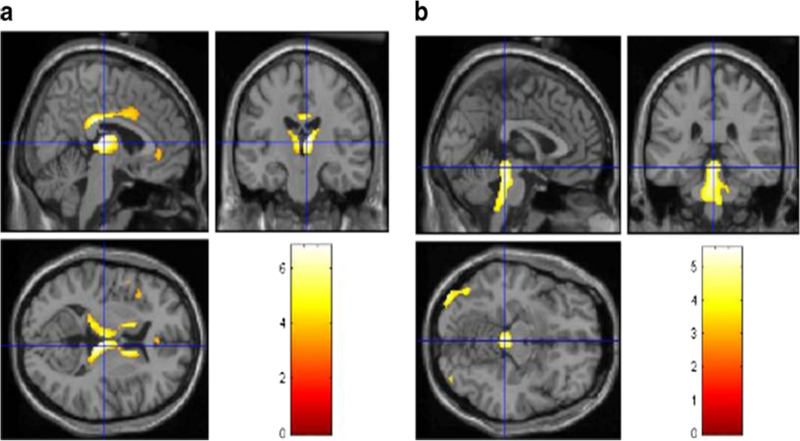
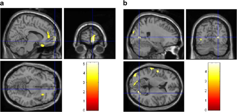
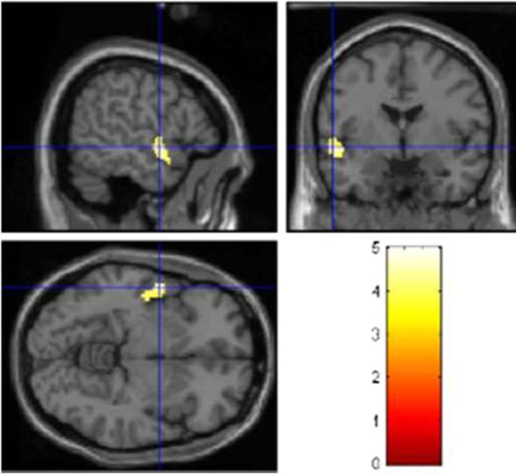
Results: The C9ORF72-ALS patients compared with the sALS patients showed significant hypometabolism in the anterior and posterior cingulate cortex, insula, caudate and thalamus, the left frontal and superior temporal cortex, and hypermetabolism in the midbrain, bilateral occipital cortex, globus pallidus and left inferior temporal cortex. The ALS-FTD patients compared with the sALS patients showed more limited hypometabolic areas, including the orbitofrontal, prefrontal, anterior cingulate and insular cortex, and hypermetabolic areas, including the bilateral occipital cortex, the left precentral and postcentral cortex and superior temporal gyrus. The C9ORF72-ALS patients compared with the ALS-FTD patients showed hypometabolism in the left temporal cortex.

Conclusion: ALS patients with the C9ORF72 hexanucleotide repeat expansion had a more widespread central nervous system involvement than ALS patients without genetic mutations, with or without comorbid FTD, consistent with their more severe clinical picture.


Figures

Fig. 1
Fig. 1
PET findings in 15 C9ORF72-ALS patients compared with 30 sALS patients. Statistically significant differences (p < 0.05 FDR) are highlighted on a MRI T1 template. a Hypometabolism: top left sagittal view,top right coronal view. bottom left transverse view. Significant corrected clusters are seen in the bilateral anterior and posterior cingulate cortex, insula, caudate and thalamus, and the left frontal and superior temporal cortices. b Hypermetabolism: top left sagittal view, top right coronal view, bottom left transverse view. Significant corrected clusters are seen in the midbrain, bilateral occipital cortex, the globus pallidus, and the left middle and inferior temporal cortices.
Fig. 2
Fig. 2
PET findings in 12 ALS-FTD patients compared with 30 sALS patients. Statistically significant differences (p < 0.001) are highlighted on a MRI T1 template. a Hypometabolism: top left sagittal view, top right coronal view, bottom left transverse view. Significant corrected clusters are seen in the right orbitofrontal, prefrontal, anterior cingulate and insular cortices. b Hypermetabolism: top left sagittal view, top right coronal view,bottom left transverse view. Significant corrected clusters are seen in the bilateral occipital cortex, in the left precentral and postcentral cortex and the superior temporal gyrus
Fig. 3
Fig. 3
PET findings in 15 C9ORF72-ALS patients compared with 12 ALS-FTD patients. Statistically significant differences (p < 0.001) are highlighted on a MRI T1 template. Hypometabolism: top left sagittal view, top right coronal view, bottom left transverse view. Significant corrected clusters are seen in the left temporal cortex. No significant hypermetabolism was found in this comparison

References

    1. Kiernan MC, Vucic S, Cheah BC, Turner MR, Eisen A, Hardiman O, et al. Amyotrophic lateral sclerosis. Lancet. 2011;377:942–55. - PubMed
    1. Byrne S, Elamin M, Bede P, Shatunov A, Walsh C, Corr B, et al. Cognitive and clinical characteristics of patients with amyotrophic lateral sclerosis carrying a C9ORF72 repeat expansion: a population-based cohort study. Lancet Neurol. 2012;11:232–40. - PMC - PubMed
    1. Chiò A, Borghero G, Restagno G, Mora G, Drepper C, Traynor BJ, et al. Clinical characteristics of patients with familial amyotrophic lateral sclerosis carrying the pathogenic GGGGCC hexanucleotide repeat expansion of C9ORF72. Brain. 2012;135:784–93. - PMC - PubMed
    1. Dejesus-Hernandez M, Mackenzie IR, Boeve BF, Boxer AL, Baker M, Rutherford NJ, et al. Expanded GGGGCC hexanucleotide repeat in noncoding region of C9ORF72 causes chromosome 9p-linked FTD and ALS. Neuron. 2011;72:245–56. - PMC - PubMed
    1. Renton AE, Majounie E, Waite A, Simón-Sánchez J, Rollinson S, Gibbs JR, et al. A hexanucleotide repeat expansion in C9ORF72 is the cause of chromosome 9p21-linked ALS-FTD. Neuron. 2011;72:257–68. - PMC - PubMed

Publication types