. 2014 Mar;39(5):744-56.
doi: 10.1111/ejn.12484. Epub 2014 Jan 22.

A reduction in Ptprq associated with specific features of the deafness phenotype of the miR-96 mutant mouse diminuendo

Affiliations
Free PMC article

A reduction in Ptprq associated with specific features of the deafness phenotype of the miR-96 mutant mouse diminuendo

Jing Chen et al. Eur J Neurosci. 2014 Mar.
Free PMC article

Abstract

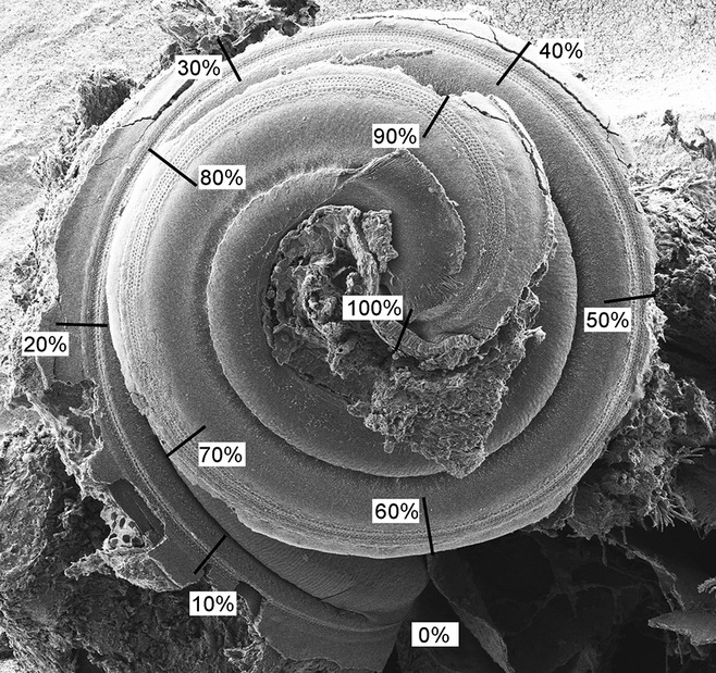
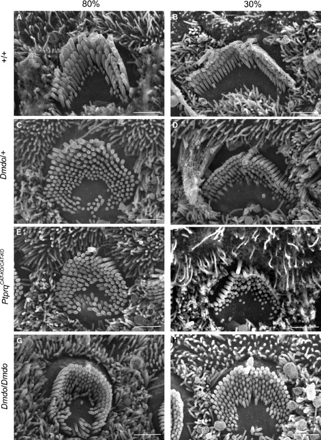
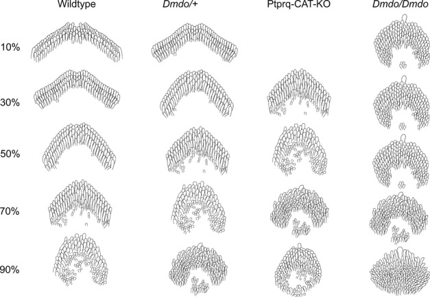
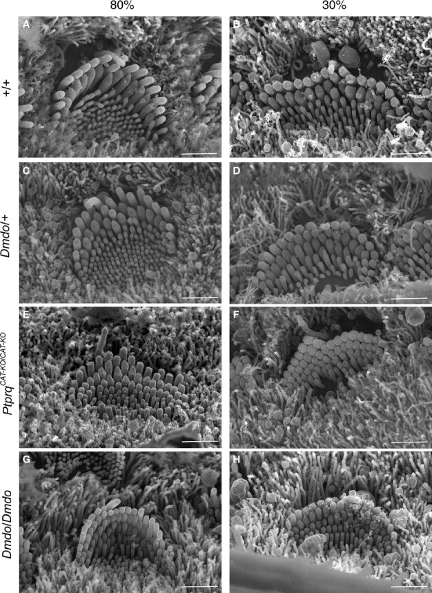
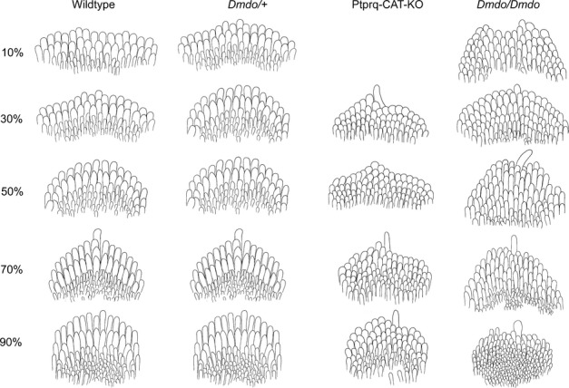
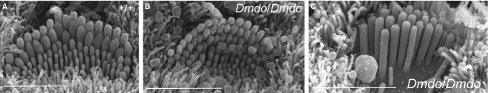
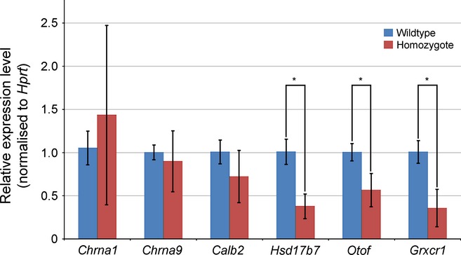
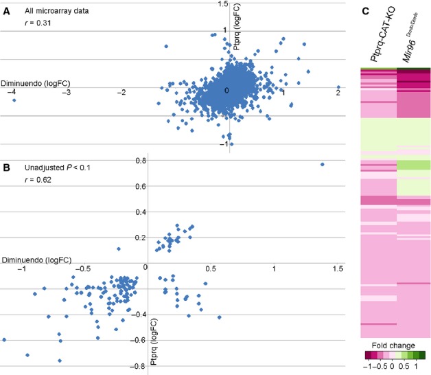
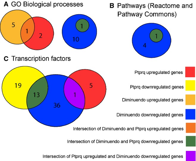
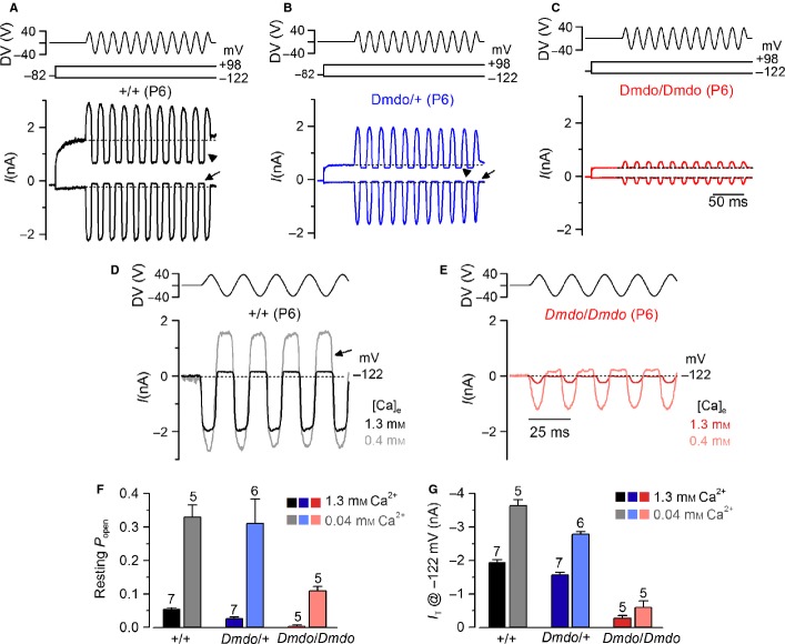
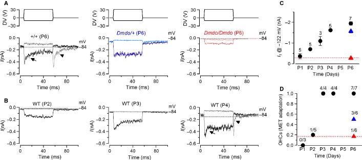
miR-96 is a microRNA, a non-coding RNA gene which regulates a wide array of downstream genes. The miR-96 mouse mutant diminuendo exhibits deafness and arrested hair cell functional and morphological differentiation. We have previously shown that several genes are markedly downregulated in the diminuendo organ of Corti; one of these is Ptprq, a gene known to be important for maturation and maintenance of hair cells. In order to study the contribution that downregulation of Ptprq makes to the diminuendo phenotype, we carried out microarrays, scanning electron microscopy and single hair cell electrophysiology to compare diminuendo mutants (heterozygous and homozygous) with mice homozygous for a functional null allele of Ptprq. In terms of both morphology and electrophysiology, the auditory phenotype of mice lacking Ptprq resembles that of diminuendo heterozygotes, while diminuendo homozygotes are more severely affected. A comparison of transcriptomes indicates there is a broad similarity between diminuendo homozygotes and Ptprq-null mice. The reduction in Ptprq observed in diminuendo mice appears to be a major contributor to the morphological, transcriptional and electrophysiological phenotype, but does not account for the complete diminuendo phenotype.

Keywords: ear development; hereditary hearing loss; knockout and transgenic m; molecular genetics; sensory hair cells.


Figures

Fig. 1
Fig. 1
Method of measuring the length of the cochlear duct to make comparisons in corresponding positions. In order to compare hair cell bundles in corresponding positions along cochlear ducts between wildtype and mutant mice or between different mutant mice, the images were taken under a standard magnification (× 80) to show the whole organ of Corti. The cochlear duct was divided into ten parts of 10% from base to apex. A higher magnification (× 15 k) was used for analysis of IHCs and the outermost row of OHCs at every 10% interval along the length of the cochlear duct. Scale bar, 300 μm.
Fig. 2
Fig. 2
Outer hair cell stereocilia morphology at position 80% (near apex; A, C, E and G) and 30% (near base; B, D, F and H) of the length along the cochlear duct (P4) by scanning electron microscopy showed developmentally immature morphology of hair bundles in the mutants. (G and H) Diminuendo homozygotes showed the most affected hair bundles, with a rounded, almost circular shape and extra stereocilia rows. (C and D) Diminuendo heterozygotes resemble (E and F) Ptprq-CAT-KO homozygotes, being less affected but still showing differences compared to (A and B) wildtypes, such as the gently rounded hair bundle and excess microvilli. Scale bar, 1.5 μm.
Fig. 3
Fig. 3
Cartoons of P4 OHCs typical of wildtype, diminuendo heterozygote, Ptprq-CAT-KO homozygote and diminuendo homozygote mice. The wildtype hair cells (left) display the typical gradient of development of outer hair cells at P4, with the most mature hair cells at the base (10%) and the most immature at the apex (90%). This gradient is still present in diminuendo heterozygote mice (centre left), but appears delayed; at each stage depicted, the hair cells resemble the next most apical stage of the wildtype. A similar difference can be observed between Ptprq-CAT-KO homozygote OHCs (centre right) and diminuendo heterozygote OHCs, with Ptprq-CAT-KO OHCs at 30% resembling diminuendo heterozygote OHCs at 50%, which resemble wildtype OHCs at 70%, for example. No Ptprq-CAT-KO OHCs were preserved for observation at 10%. Diminuendo homozygotes (right) display a much more extreme phenotype, with hair cell development appearing to stall at ∼ 50%, and hair cells more apical than that appearing very immature.
Fig. 4
Fig. 4
Inner hair cell stereocilia morphology at position 80% (near apex; A, C, E and G) and 30% (near base; B, D, F and H) along the length of the cochlear duct (P4) by scanning electron microscopy showed developmentally immature morphology of hair bundles in the mutants. (G and H) Diminuendo homozygote hair bundles show very little thickening of stereocilia rows. Like the outer hair cells, (C and D) diminuendo heterozygotes and (E and F) Ptprq-CAT-KO homozygotes show a less extreme phenotype but are still distinct from (A and B) the wildtype hair bundles, with an abundance of microvilli. Scale bar, 1.5 μm.
Fig. 5
Fig. 5
Cartoons of P4 IHCs typical of wildtype, diminuendo heterozygote, Ptprq-CAT-KO homozygote and diminuendo homozygote mice. Wildtype IHCs (left) show a gradient of maturity from the base (most mature, 10%) to the apex (least mature, 90%). Diminuendo heterozygotes (centre left) display a very similar gradient, with a slight delay in development visible from 10 to 30%. Ptprq-CAT-KO homozygote IHCs (centre right) are more affected, although stereocilia are organised into rows, and the taller stereocilia show some thickening as in wildtype and diminuendo heterozygote IHCs. No Ptprq-CAT-KO IHCs were preserved for observation at 10%. Diminuendo homozygote IHCs (right) are much more affected than the others, with extra rows of stereocilia and not much thickening of the taller stereocilia, but they still appear to have a gradient of maturity between 10 and 90%.
Fig. 6
Fig. 6
Two distinct stereocilia bundle phenotypes were observed in diminuendo homozygote inner hair cells. (A) A wildtype hair cell at 40%, showing the development of the ‘staircase’ of stereocilia heights. Microvilli are still present. (B) This is the more common diminuendo homozygote phenotype. The diminuendo homozygote hair cell resembles the wildtype but appears less mature, with still-thin stereocilia and more microvilli. (C) In contrast, a minority of homozygote inner hair cells lacked the staircase organisation and microvilli entirely. We observed no hair bundles intermediate between these two phenotypes. Scale bars, 300 μm.
Fig. 7
Fig. 7
Quantitative real time PCR on cDNA from organ of Corti RNA from Ptprq-CAT-KO homozygote and wildtype littermates at P4. Three pairs were used for each comparison. Quantities have been normalised to Hprt levels, and Jag1 was used to control for sensory tissue; all pairs used showed no significant difference in expression of Jag1. Hsd17b7, Otof and Grxcr1 are all significantly downregulated in Ptprq-CAT-KO homozygotes. *P < 0.01. Error bars represent SD.
Fig. 8
Fig. 8
(A and B) Scattergraphs showing correlation of data from the two microarrays; A shows the correlation of all genes present in both microarrays regardless of significance while B shows the correlation of those genes with an unadjusted P < 0.1 in both microarrays. (C) Heatmap created using the R statistical software package, showing genes with an unadjusted P < 0.1 from the Ptprq-CAT-KO microarray (left) compared with the same genes (also with unadjusted P < 0.1) from the diminuendo microarray (right). Genes are compared and coloured according to their proportional change, on a scale from dark pink (most downregulated) to dark green (most upregulated).
Fig. 9
Fig. 9
Venn diagrams showing the numbers of gene sets significantly enriched in the diminuendo and Ptprq microarrays, and how many are common to both. (A) Gene sets annotated with GO biological processes (from the mSigDB dataset); (B) gene sets defined by Pathway Commons and Reactome corresponding to known biological pathways; (C) gene sets regulated by transcription factors (from the mSigDB dataset).
Fig. 10
Fig. 10
Mechanotransducer currents in diminuendo cochlear outer hair cells. (A–C) Saturating transducer currents recorded from (A) a control (B), a heterozygous and (C) a homozygous mutant P6 apical-coil diminuendo OHC by applying sinusoidal force stimuli of 50 Hz to the hair bundles at −122 mV and + 98 mV. The driver voltage (DV) signal of ± 40 V to the fluid jet is shown above the traces (negative deflections of the DV are inhibitory). The holding potential was −82 mV. Extracellular Ca2+ concentration was 1.3 mm. The arrows and arrowheads indicate the closure of the transducer currents (i.e. resting current) elicited during inhibitory bundle displacements at hyperpolarized and depolarized membrane potentials, respectively. Note that the resting current increases with membrane depolarization. Dashed lines indicate the holding current, which is the current at the holding membrane potential. (D and E) Comparison of transducer currents recorded from (D) a control and (E) a homozygous mutant P6 diminuendo OHC in the presence of 1.3 mm Ca2+ (black/red) and endolymphatic-like Ca2+ concentration (0.04 mm; grey/pink lines) at −122 mV. (F and G) Resting current (F) and (G) peak transducer current at a membrane potential of −122 mV recorded in OHCs from the three genotypes in the presence of 1.3 mm (black/blue/red) and 0.04 mm (grey/pale blue/pink) extracellular Ca2+.
Fig. 11
Fig. 11
The development of transducer currents in diminuendo mutant OHCs is prematurely arrested. (A) Driver voltages to the fluid jet (top panels) and transducer currents recorded at −84 mV from a control, a heterozygous and a homozygous diminuendo mutant P6 OHC. Positive driver voltages (excitatory direction) elicited inward (negative) transducer currents that declined or adapted over time only in control OHCs (arrow). A small transducer current was present at rest (before t = 0) and inhibitory bundle displacement turned this off. Upon termination of the inhibitory stimulus, the transducer current in control OHCs showed evidence of rebound adaptation (arrowhead). (B) Example of transducer currents in developing control OHCs (P2, P3 and P4). Note that both signs of current adaptation (arrow and arrowhead) are first detected at P4. (C) Maximal transducer current recorded at different postnatal ages in control OHCs (black) and that of the heterozygous (blue) and homozygous (red) diminuendo mutant cells at P6. The number of OHCs tested is shown above each data point. (D) Extent of adaptation for small bundle deflection towards the excitatory directions (see arrows in panel A, left panel) of the transducer current at different postnatal ages in control OHCs (black) and that of the heterozygous (blue) and homozygous (red) diminuendo mutant cells at P6. The number of OHCs showing MET current adaptation over the total number of cells tested is shown above each data point. Note that (C) the size and (D) the extent of adaptation recorded in P6 homozygous diminuendo OHCs is similar to that measured in P1–2 control cells.

References

    1. Benjamini Y, Hochberg Y. Controlling the false discovery rate: a practical and powerful approach to multiple testing. J. R. Stat. Soc. Ser. B. 1995;57:289–300.
    1. Bolstad B. 2001. Probe level quantile normalization of high density oligonucleotide array data. Available at http://bmbolstad.com/stuff/qnorm.pdf.
    1. Bolstad BM, Irizarry RA, Astrand M, Speed TP. A comparison of normalization methods for high density oligonucleotide array data based on variance and bias. Bioinformatics. 2003;19:185–193. - PubMed
    1. Bosher SK, Warren RL. Very low calcium content of cochlear endolymph, an extracellular fluid. Nature. 1978;273:377–378. - PubMed
    1. Chen J, Ingham N, Clare S, Raisen C, Vancollie VE, Ismail O, McIntyre RE, Tsang SH, Mahajan VB, Dougan G, Adams DJ, White JK, Steel KP. Mcph1-deficient mice reveal a role for MCPH1 in otitis media. PLoS ONE. 2013;8:e58156. - PMC - PubMed

Publication types

MeSH terms

Substances