

Recovery of neurological function of ischemic stroke by application of conditioned medium of bone marrow mesenchymal stem cells derived from normal and cerebral ischemia rats
- PMID: 24447306
- PMCID: PMC3922747
- DOI: 10.1186/1423-0127-21-5
Recovery of neurological function of ischemic stroke by application of conditioned medium of bone marrow mesenchymal stem cells derived from normal and cerebral ischemia rats
Abstract
Background: Several lines of evidence have demonstrated that bone marrow-derived mesenchymal stem cells (BM-MSC) release bioactive factors and provide neuroprotection for CNS injury. However, it remains elusive whether BM-MSC derived from healthy donors or stroke patients provides equal therapeutic potential. The present work aims to characterize BM-MSC prepared from normal healthy rats (NormBM-MSC) and cerebral ischemia rats (IschBM-MSC), and examine the effects of their conditioned medium (Cm) on ischemic stroke animal model.
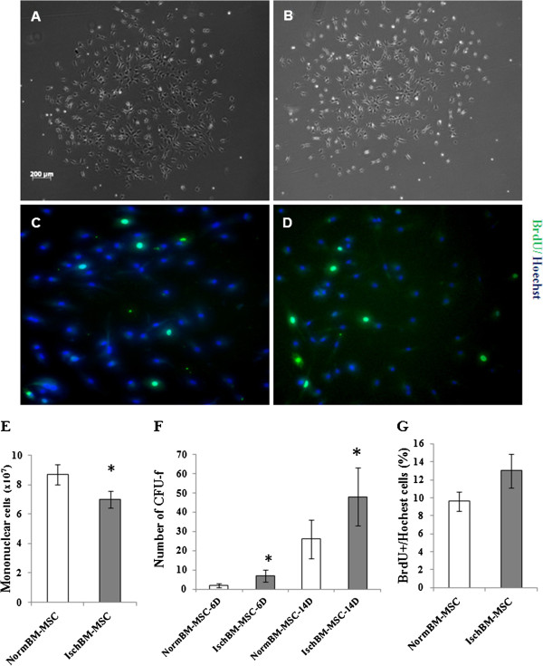
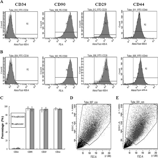
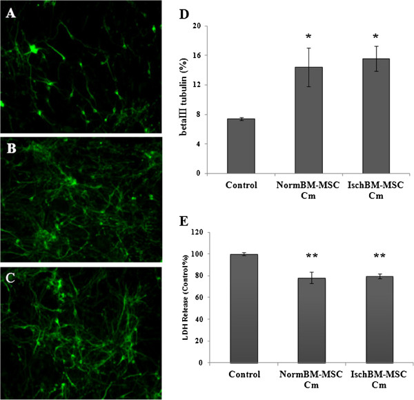
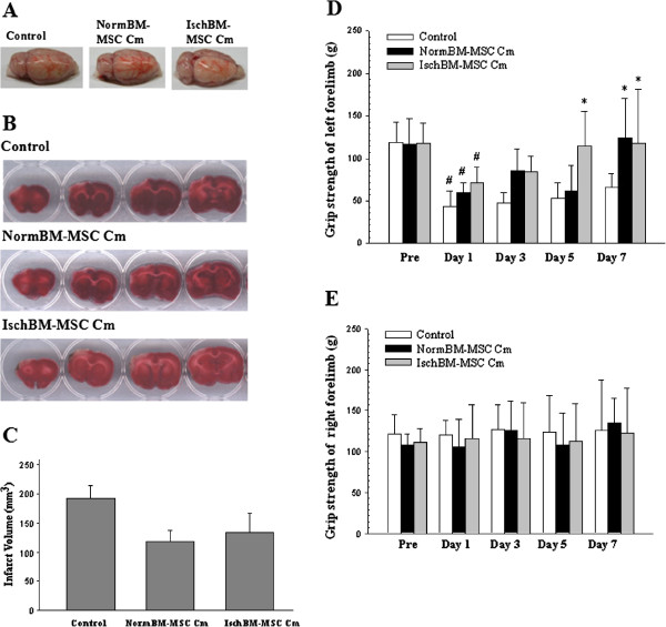
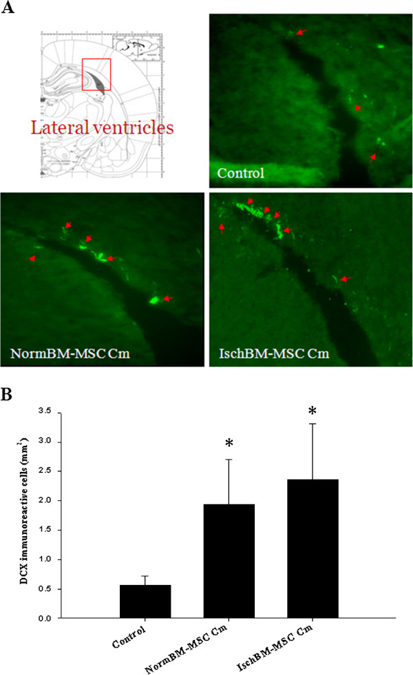
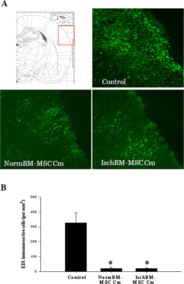
Results: Isolated NormBM-MSC or IschBM-MSC formed fibroblastic like morphology and expressed CD29, CD90 and CD44 but failed to express the hematopoietic marker CD34. The number of colony formation of BM-MSC was more abundant in IschBM-MSC than in NormBM-MSC. This is in contrast to the amount of Ficoll-fractionated mononuclear cells from normal donor and ischemic rats. The effect of cm of BM-MSC was further examined in cultures and in middle cerebral artery occlusion (MCAo) animal model. Both NormBM-MSC Cm and IschBM-MSC Cm effectively increased neuronal connection and survival in mixed neuron-glial cultures. In vivo, intravenous infusion of NormBM-MSC Cm and IschBM-MSC Cm after stroke onset remarkably improved functional recovery. Furthermore, NormBM-MSC Cm and IschBM-MSC Cm increased neurogenesis and attenuated microglia/ macrophage infiltration in MCAo rat brains.
Conclusions: Our data suggest equal effectiveness of BM-MSC Cm derived from ischemic animals or from a normal population. Our results thus revealed the potential of BM-MSC Cm on treatment of ischemic stroke.
Figures
Similar articles
-
Minocycline exhibits synergism with conditioned medium of bone marrow mesenchymal stem cells against ischemic stroke.J Tissue Eng Regen Med. 2021 Mar;15(3):279-292. doi: 10.1002/term.3171. Epub 2021 Feb 9. J Tissue Eng Regen Med. 2021. PMID: 33470523
-
A comparative study of different doses of bone marrow-derived mesenchymal stem cells improve post-stroke neurological outcomes via intravenous transplantation.Brain Res. 2023 Jan 1;1798:148161. doi: 10.1016/j.brainres.2022.148161. Epub 2022 Nov 12. Brain Res. 2023. PMID: 36379315
-
Serum-mediated Activation of Bone Marrow-derived Mesenchymal Stem Cells in Ischemic Stroke Patients: A Novel Preconditioning Method.Cell Transplant. 2018 Mar;27(3):485-500. doi: 10.1177/0963689718755404. Epub 2018 May 18. Cell Transplant. 2018. PMID: 29774769 Free PMC article.
-
Neuroprotection by mesenchymal stem cell (MSC) administration is enhanced by local cooling infusion (LCI) in ischemia.Brain Res. 2019 Dec 1;1724:146406. doi: 10.1016/j.brainres.2019.146406. Epub 2019 Aug 24. Brain Res. 2019. PMID: 31454517 Review.
-
Evidence for high translational potential of mesenchymal stromal cell therapy to improve recovery from ischemic stroke.J Cereb Blood Flow Metab. 2013 Sep;33(9):1322-34. doi: 10.1038/jcbfm.2013.91. Epub 2013 Jun 12. J Cereb Blood Flow Metab. 2013. PMID: 23756689 Free PMC article. Review.
Cited by
-
Secretome as a Tool to Treat Neurological Conditions: Are We Ready?Int J Mol Sci. 2023 Nov 20;24(22):16544. doi: 10.3390/ijms242216544. Int J Mol Sci. 2023. PMID: 38003733 Free PMC article. Review.
-
Effect of exosomes derived from multipluripotent mesenchymal stromal cells on functional recovery and neurovascular plasticity in rats after traumatic brain injury.J Neurosurg. 2015 Apr;122(4):856-67. doi: 10.3171/2014.11.JNS14770. Epub 2015 Jan 16. J Neurosurg. 2015. PMID: 25594326 Free PMC article.
-
Recent Advances in Mono- and Combined Stem Cell Therapies of Stroke in Animal Models and Humans.Int J Mol Sci. 2019 Nov 29;20(23):6029. doi: 10.3390/ijms20236029. Int J Mol Sci. 2019. PMID: 31795466 Free PMC article. Review.
-
Human neural stem cell secretome relieves endoplasmic reticulum stress-induced apoptosis and improves neuronal functions after traumatic brain injury in a rat model.J Mol Histol. 2024 Jun;55(3):329-348. doi: 10.1007/s10735-024-10192-7. Epub 2024 Apr 12. J Mol Histol. 2024. PMID: 38609527
-
Systemic administration of cell-free exosomes generated by human bone marrow derived mesenchymal stem cells cultured under 2D and 3D conditions improves functional recovery in rats after traumatic brain injury.Neurochem Int. 2017 Dec;111:69-81. doi: 10.1016/j.neuint.2016.08.003. Epub 2016 Aug 15. Neurochem Int. 2017. PMID: 27539657 Free PMC article.
References
Publication types
MeSH terms
Substances
LinkOut - more resources
Full Text Sources
Other Literature Sources
Medical
Miscellaneous
