. 2014 Jan 21:9:16.
doi: 10.1186/1746-1596-9-16.

Expression of serum amyloid A in uterine cervical cancer

Affiliations

Expression of serum amyloid A in uterine cervical cancer

Yanjie Ren et al. Diagn Pathol. .

Abstract

Background: As an acute-phase protein, serum amyloid A (SAA) is expressed primarily in the liver. However, its expression in extrahepatic tissues, especially in tumor tissues, was also demonstrated recently. In our study, we investigated the expression of SAA in uterine cervical carcinomas, and our results suggested its potential as a serum biomarker.

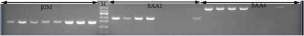
Methods: Quantitative real-time polymerase chain reaction (RT-PCR), immunohistochemistry (IHC) and enzyme-linked immunosorbent assay (ELISA) were used to evaluate the SAA gene and protein expression levels in the tissues and sera of patients with non-neoplastic lesions (NNLs), cervical intraepithelial neoplasia (CIN) and cervical carcinoma (CC).

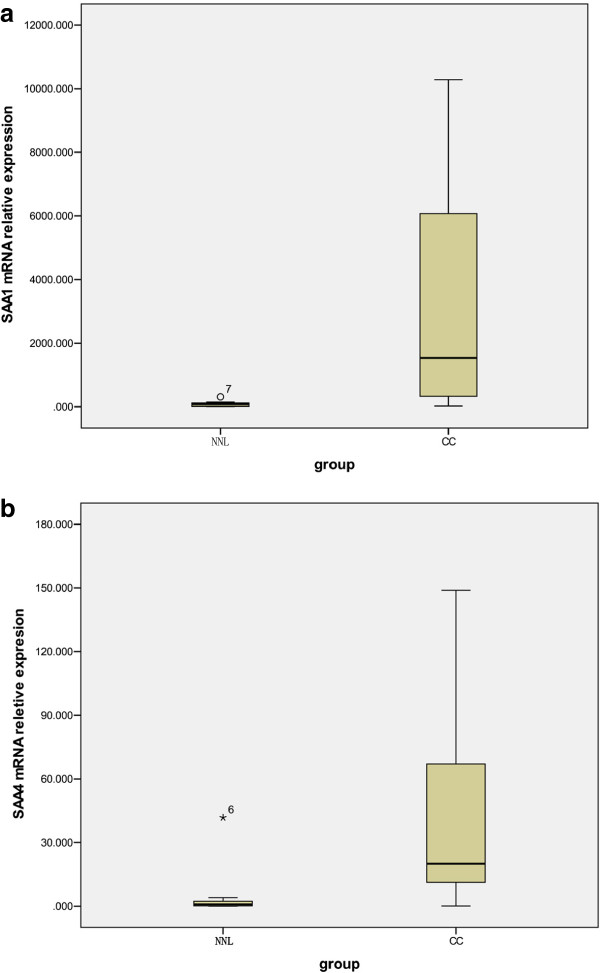
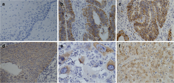
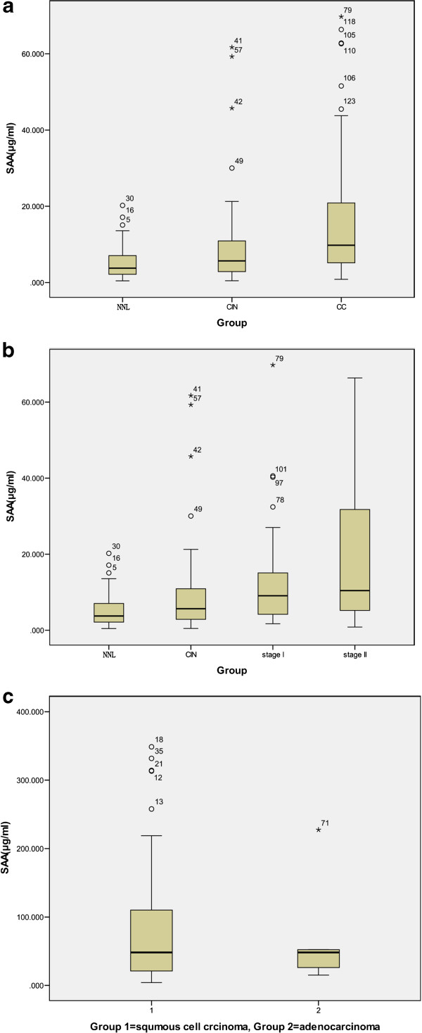
Results: Compared with NNLs, the SAA gene (SAA1 and SAA4) expression levels were significantly higher in uterine CC (mean copy numbers: 138.7 vs. 5.01, P < 0.000; and 1.8 vs. 0.079, P = 0.001, respectively) by real-time PCR. IHC revealed cytoplasmic SAA protein staining in tissues from adenocarcinoma and squamous cell carcinoma of the cervix. The median serum concentrations (μg/ml) of SAA were 6.02 in patients with NNLs and 10.98 in patients with CIN (P = 0.31). In contrast, the median serum SAA concentration was 23.7 μg/ml in uterine CC patients, which was significantly higher than the SAA concentrations of the NNL group (P = 0.002) and the CIN group (P = 0.024).

Conclusions: Our data suggested that SAA might be a uterine CC cell product. High SAA concentrations in the serum of CC patients may have a role in monitoring disease occurrence and could have therapeutic applications.

Virtual slides: The virtual slide(s) for this article can be found here: http://www.diagnosticpathology.diagnomx.eu/vs/1433263219102962.


Figures

Figure 1
Figure 1
mRNA expression of SAA1 and SAA4 in freshly frozen biopsies. Expression of SAA1 (a) and SAA4 (b) by quantitative real-time polymerase chain reaction (RT-PCR) in 10 non neoplastic lesion cervical control samples and 21 cervical carcinoma freshly frozen biopsies are shown. The vertical axis represents the relative values of threshold cycle corrected with β2M (2-ΔCt). Values are means ± SD of triplicate measurement.
Figure 2
Figure 2
SAA mRNA expression by electrophoresis. Representive SAA1 and SAA4 PCR fragments were analyzed on a 2% agarose gel. In each 8 lanes, the first four were derived from different cervical carcinoma tissues and the rest were from non neoplastic lesion cervical tissues. Markers of a DNA ladder (50-bp steps) are shown in lane M. Using primers for SAA1, SAA4, and the control gene β-2 M analysis 10 benign cervical and 21 cervical carcinomas (2 adenocarcinomas and 19 squamous cell carcinomas) patients by RT-PCR.
Figure 3
Figure 3
SAA protein expression by IHC. IHC demonstrating SAA protein expression in cervical carcinomas. The sections were immunostained with monoclonal anti-SAA antibodies. The reddish-brown staining represents positive SAA protein signal; counterstaining is light blue. (a) Non neoplastic lesion cervical tissue. The tumoral epithelium stained moderate to strong and focal. (b, c) Adenocarcinoma. The stage I cervical squamous carcinoma tissues stained moderate and focal. (d) and diffuse (e).Stage II. Strong cytoplasmic SAA positivity was evident in the positive control (f) Liver. Original magnification x400 (a-f).
Figure 4
Figure 4
SAA serum levels by ELISA. (a) SAA serum levels in patients with cervical carcinoma. Serum levels of SAA were measured in patients with cervical pathologies classified as follows: (1) Non neoplastic lesion gynecologic diseases (n32), (2) cervical intraepithelial dysplasia (n35), (3) cervical carcinoma (n72). (b) SAA levels in cervical carcinoma with different degrees of stage (ie, 34 stages I, 37 stages II) are shown. Data are presented as mean ± standard deviation of mean. (c) SAA levels in cervical carcinoma with different histological types (ie, 66 squamous cell carcinomas, 6 adenocarcinomas) are shown.

References

    1. Jemal A, Bray F, Center MM, Ferlay J, Ward E, Forman D. Global cancer statistics. CA Cancer J Clin. 2011;61:69–90. doi: 10.3322/caac.20107. - DOI - PubMed
    1. Franco E, Monsonego J. New Developments in Cervical Cancer Screening and Prevention. Oxford: Blackwell Science Press; 1997.
    1. Morris M, Tortolero-Luna G, Malpica A, Baker VV, Cook E, Johnson E, Mitchell MF. Cervical intraepithelial neoplasia and cervical cancer. Obstet Gynecol Clin North Am. 1996;23:347–410. - PubMed
    1. Ponten J, Adami HO, Bergstro MR, Dillner J, Friberg LG, Gustafsson L, Miller AB, Parkin DM, Sparen P, Trichopoulos D. Strategies for global control of cervical cancer. Int J Cancer. 1995;60:1–26. doi: 10.1002/ijc.2910600102. - DOI - PubMed
    1. Cocco E, Bellone S, El-Sahwi K, Cargnelutti M, Buza N, Tavassoli FA, Schwartz PE, Thomas J, Rutherford, Pecorelli S, Santin AD. Serum amyloid A: a novel biomarker for endometrial cancer. Cancer. 2010;116:843–851. doi: 10.1002/cncr.24838. - DOI - PMC - PubMed

Publication types

MeSH terms