. 2014 May;26(5):933-41.
doi: 10.1016/j.cellsig.2014.01.005. Epub 2014 Jan 19.

Docosahexaenoic acid reverses angiotensin II-induced RECK suppression and cardiac fibroblast migration

Affiliations

Docosahexaenoic acid reverses angiotensin II-induced RECK suppression and cardiac fibroblast migration

Jalahalli M Siddesha et al. Cell Signal. 2014 May.

Abstract

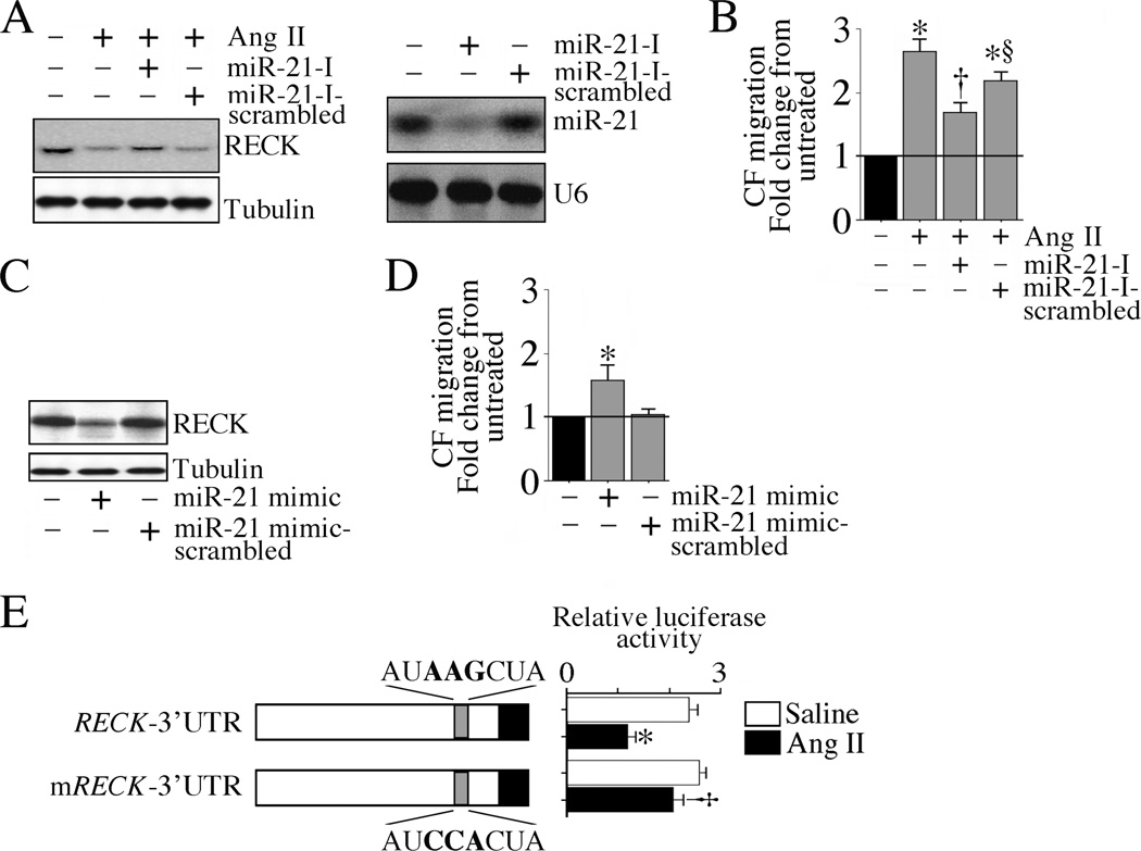
The omega-3 polyunsaturated fatty acids (ω-3 fatty acids) eicosapentaenoic acid (EPA) and docosahexaenoic acid (DHA) have been reported to inhibit or delay the progression of cardiovascular diseases, including myocardial fibrosis. Recently we reported that angiotensin II (Ang II) promotes cardiac fibroblast (CF) migration by suppressing the MMP regulator reversion-inducing-cysteine-rich protein with Kazal motifs (RECK), through a mechanism dependent on AT1, ERK, and Sp1. Here we investigated the role of miR-21 in Ang II-mediated RECK suppression, and determined whether the ω-3 fatty acids reverse these effects. Ang II induced miR-21 expression in primary mouse cardiac fibroblasts (CFs) via ERK-dependent AP-1 and STAT3 activation, and while a miR-21 inhibitor reversed Ang II-induced RECK suppression, a miR-21 mimic inhibited both RECK expression and Ang II-induced CF migration. Moreover, Ang II suppressed the pro-apoptotic PTEN, and the ERK negative regulator Sprouty homologue 1 (SPRY1), but induced the metalloendopeptidase MMP2, all in a manner that was miR-21-dependent. Further, forced expression of PTEN inhibited Akt phosphorylation, Sp1 activation, and MMP2 induction. Notably, while both EPA and DHA reversed Ang II-mediated RECK suppression, DHA appeared to be more effective, and reversed Ang II-induced miR-21 expression, RECK suppression, MMP2 induction, and CF migration. These results indicate that Ang II-induced CF migration is differentially regulated by miR-21-mediated MMP induction and RECK suppression, and that DHA has the potential to upregulate RECK, and therefore may exert potential beneficial effects in cardiac fibrosis.

Keywords: Fibrosis; MicroRNA; PTEN; RECK; SPRY1; ω−3 lipids.


Conflict of interest statement

Conflict of Interest: None.

Figures

Fig. 1
Fig. 1. ω-3 polyunsaturated fatty acids reverse Ang II-induced miR-21 expression and RECK suppression
A, EPA and DHA upregulate basal RECK expression. At 50-70% confluency, adult mouse cardiac fibroblasts (CF) were made quiescent by incubating in medium containing 0.5% BSA (serum-free) for 48 h. The quiescent CF were then treated with EPA or DHA (100 µM) for 8 h. RECK expression levels were analyzed by immunoblotting. Densitometric analysis of immunoreactive bands from three independent experiments is summarized in the adjacent panel. *P < at least 0.05 vs. untreated (n=3). B, EPA and DHA reverse Ang II-induced RECK suppression. The quiescent CF were treated with EPA or DHA (100 µM for 8 h) prior to Ang II addition (10−7M for 6 h). RECK expression was analyzed as in A, and the densitometric analysis is shown on the right. *P < 0.05 vs. untreated, †P < 0.05 vs. Ang II (n=3). C, Both DHA and forced expression of RECK attenuate Ang II-induced CF migration. CF infected with Ad.RECK (moi 40) for 24 h, or pretreated with DHA as B, were layered on Matrigel™ basement membrane matrix-coated filters, and then treated with Ang II (10−7M for 12 h). The lower chamber contained medium with 10% serum. Cells migrating to the other side of the membrane were quantified using MTT assay. C, *P < 0.01 vs. untreated, †P < 0.05 vs. Ang II, § P < 0.01 vs. Ang II+DHA (n=6).
Fig. 2
Fig. 2. Ang II induces CF migration via miR-21-dependent RECK suppression
A, miR-21 inhibitor reverses Ang II-induced RECK suppression. CF transfected with miR-21 inhibitor (miR-21-I; 80 nM for 24 h) were treated with Ang II (10−7M) for 6 h, and then analyzed for RECK expression by immunoblotting. Inhibition in miR-21 expression was confirmed by Northern blotting as shown on the right (n=3). B, miR-21 inhibitor reverses Ang II-induced CF migration. CF transfected with the miR-21 inhibitor (80 nM for 24 h) were layered on Matrigel™ basement membrane matrix-coated filters, incubated with Ang II (10−7M) for 12 h, and then analyzed for migration by MTT assay. *P < 0.01 vs. untreated; †P < 0.05 vs. Ang II, § P < 0.05 vs. Ang II + miR-21-I (n=6). C, miR-21 mimic inhibits basal RECK expression. CF transfected with miR-21 mimic or its scrambled control (80 nM for 24 h) were analyzed for RECK expression by immunoblotting (n=3). D, miR-21 mimic stimulates CF migration. CF transfected with miR-21 mimic (80 nM) for 24 h were analyzed for CF migration as in B. *P < 0.05 vs. untreated (n=6). E, Targeting miR-21 reverses Ang II-mediated RECK suppression. CF transfected with wild type (pGL3-RECK-3’UTR) or miR-21 mutated (pGL3-mRECK-3’UTR) RECK 3’UTR reporter vector (2 µg) along with pRL-Tk vector (100 ng) for 24 h were treated with Ang II (10−7M for 12 h), and then harvested for dual luciferase activity. *P < 0.05 vs. Saline-untreated, †P < 0.01 vs. mutant RECK (n=6).
Fig. 3
Fig. 3. DHA reverses Ang II-induced miR-21 expression
A, Ang II induces miR-21 via ERK. The quiescent CF were treated with PD98059 (10 µM) for 1 h prior to Ang II addition (10−7M for 1 h). miR-21 expression was analyzed by RT-qPCR and Northern blotting (inset). U6 snRNA served as a loading control (n=3). B, Ang II-induced miR-21 expression is AP-1 dependent. CF transduced with lentiviral c-Jun shRNA (moi 0.5 for 48 h) were incubated with Ang II (10−7M for 1 h). miR-21 expression was analyzed as in A (n=3). C, Ang II-induced MIR21 promoter-reporter activity is AP-1-dependent. CF transfected with miPPR-21-410 promoter reporter or its deletion constructs (2 μg) along with the Renilla luciferase vector (pRL-TK, 100 ng) for 24 h were treated with Ang II (10− 7 M for 12 h), and harvested for the dual-luciferase assay. D, DHA inhibits Ang II-induced miR-21 promoter reporter activity. CF were transfected with miPPR-21-410 promoter reporter (2 μg) along with pRL-Tk (100 ng) for 24 h, incubated with DHA (100 µM for 8 h) and then treated with Ang II (10− 7 M) for 12 h prior to harvesting for the dual-luciferase assay. E, DHA suppresses Ang II-induced miR-21 induction. The quiescent CF were treated with DHA (100 µM for 8 h) prior to Ang II addition. miR-21 expression was analyzed as in A. A, B, D, E, *P < at least 0.05 vs. untreated, †P < 0.05 vs. Ang II (n=3-6).
Fig. 4
Fig. 4. DHA inhibits Ang II-induced PTEN/Akt-dependent MMP2 expression
A, Ang II induces CF migration in part via MMP2. CF transduced with adenoviral MMP2 siRNA (moi 100 for 24 h) were layered on matrix-coated filters, incubated with Ang II (10−7 M for 12 h), and then analyzed for migration as in Fig. 2B. Knockdown of MMP2 was confirmed by immunoblotting as shown on the right (n=3). MMP9 served as an off-target. *P < 0.01 vs. untreated; †P < 0.01 vs. Ang II (n=6). B, Forced expression of PTEN or dnAkt attenuates Ang II-induced MMP2 expression. CF transduced with Ad.PTEN (moi 20) or Ad.dnAkt (moi 100) for 24 h were incubated with Ang II (10−7 M) for 1 h (mRNA expression) or 24 h (immunoblotting). MMP2 mRNA expression was analyzed by RT-qPCR and MMP2 levels in culture supernatants by immunoblotting using antibodies that detect both pro and active forms (inset; n=3). *P < 0.01 vs. untreated; †P < 0.05 vs. Ang II ± Ad.GFP (n=6). C, DHA reverses Ang II-induced PTEN suppression and Akt activation. The quiescent CF were treated with DHA (100 mM for 8 h) prior to Ang II addition (10−7M for 2 h). PTEN and p-Akt expressions were analyzed by immunoblotting (n=3). D, Ang II-induced MMP2 expression and activation is Sp1-dependent. CF transduced with lentiviral Sp1 shRNA (moi 0.5) for 48 h prior to Ang II addition (10−7 M) for either 1 (mRNA expression) or 24 h (immunoblotting) were analyzed for MMP2 mRNA expression by RT-qPCR and activity by immunoblotting using equal amounts of culture supernatants and antibodies that detect both pro and active forms (inset; n=3). Knockdown of Sp1 was confirmed by immunoblotting and is shown on the right. *P < 0.01 vs. untreated; †P < 0.05 vs. Ang II ± GFP (n=6). E, Forced expression of PTEN attenuates Ang II-induced Sp1 activation via PI3K and Akt. CF transduced with Ad.PTEN (moi 20 for 24 h) prior to Ang II addition were analyzed for phospho-Sp1 levels (Thr453) by immunoblotting using nuclear protein extracts and antibodies that detect activation-specific antibodies (n=3). Lamin A/C and GAPDH served as purity and loading controls. F, Ang II induces Sp1 activation via PI3K and Akt. The quiescent CF were treated with wortmannin or SH-5 as in A and B prior to Ang II addition. Phospho-Sp1 levels were analyzed by immunoblotting as in E (n=3). G, DHA inhibits Ang II-induced Sp1 activation. The quiescent CF were incubated with DHA (100 µM for 8 h) prior to Ang II addition (10−7 M). Phospho-Sp1 levels were analyzed as in E (n=3).
Fig. 5
Fig. 5. Sprouty homologue 1 (SPRY1) regulates RECK expression
A, miR-21 inhibitor reverses Ang II-induced SPRY1 suppression. CF transfected with miR-21 inhibitor (miR-21-I; 80 nM) for 24 h were incubated with Ang II (10−7 M) for 1 h, and then analyzed for SPRY1 expression by immunoblotting using cleared whole cell lysates (n=3). miR-21 scrambled inhibitor served as a control. B, miR-21 mimic inhibits basal SPRY1 expression. CF transfected with miR-21 mimic (80 nM) for 24 h were analyzed for SPRY1 expression as in A (n=3). C, Forced expression of SPRY1 reverses Ang II-induced ERK activation. CF transduced with Ad.SPRY1 (moi100) for 24 h were incubated with Ang II (10−7 M for 1 h). Phospho-ERK levels were analyzed by immunoblotting (n=3). SPRY1 overexpression was confirmed by immunoblotting as shown on the right. D, Forced expression of SPRY1 attenuates Ang II-induced RECK suppression. CF treated as in C, but for 6 h with Ang II (10−7 M) were analyzed for RECK expression by immunoblotting. Densitometric analysis of immunoreactive bands from three independent experiments is summarized on the right. *P < 0.05 vs. untreated; †P < 0.05 vs. Ang II (n=3).
Fig. 6
Fig. 6. DHA inhibits Ang II-induced STAT3-mediated miR-21 expression and CF migration
A, Ang II induces STAT3 phosphorylation via ERK. The quiescent CF were incubated with PD98059 (10 µM in DMSO for 1 h) prior to Ang II addition (10−7 M for 15 min). Total and phospho-STAT3 levels were analyzed by immunoblotting (n=3). B, Forced expression of mutant STAT3 attenuates Ang II-induced STAT3 phosphorylation. CF transduced with lentiviral dominant negative STAT3 (moi 0.5 for 48 h) were treated with Ang II (10−7 M) for 15 min, and then analyzed for phospho-STAT3 levels as in A (n=3). C, S31-201 inhibits Ang II-induced STAT3 phosphorylation. The quiescent CF treated with Ang II (10−7 M) and S31-201 (10 µM) simultaneously were analyzed for total and phospho-STAT3 levels as in A (n=3). D, Targeting STAT3 attenuates Ang II-induced miR-21 expression. CF treated as in B and C, but for 1 h with Ang II (10−7 M) were analyzed for miR-21 expression by RT-qPCR. E, STAT3 inhibition attenuates Ang II-induced MMP2 expression. The quiescent CF treated with S31-201 and Ang II simultaneously for 2 h were analyzed for MMP2 mRNA expression by RT-qPCR. F, STAT3 inhibition attenuates Ang II-induced CF migration. The quiescent CF layered on matrix-coated filters were incubated with Ang II (10−7 M) and S31-201 (10 µM) together for 12 h were analyzed for migration as in Fig. 2B. G, DHA inhibits Ang II-induced STAT3 phosphorylation. The quiescent CF incubated with DHA (100 µM for 8 h) prior to Ang II addition (10−7 M for 15 min) were analyzed for total and phospho-STAT3 levels as in A (n=3). D, E, F, *P < 0.01 vs. untreated; †P < at least 0.05 vs. Ang II (n=6).
Fig. 7
Fig. 7
Schema showing possible signal transduction pathways involved in Angiotensin II-induced miR-21 induction, RECK, SPRY1 and PTEN inhibition, MMP2 expression, and cardiac fibroblast migration.

Similar articles

Cited by

References

    1. Frangogiannis NG. Physiol Rev. 2012;92(2):635–688. - PMC - PubMed
    1. Shinde AV, Frangogiannis NG. J Mol Cell Cardiol. 2013 - PMC - PubMed
    1. Baker AH, Edwards DR, Murphy G. J Cell Sci. 2002;115(Pt 19):3719–3727. - PubMed
    1. Mishra PK, Givvimani S, Chavali V, Tyagi SC. Biochim Biophys Acta. 2013;1832(12):2271–2276. - PMC - PubMed
    1. Spinale FG, Janicki JS, Zile MR. Circ Res. 2013;112(1):195–208. - PMC - PubMed

Publication types

MeSH terms