. 2014 Mar 15;74(6):1718-27.
doi: 10.1158/0008-5472.CAN-13-2050. Epub 2014 Jan 21.

Neuroplastic changes occur early in the development of pancreatic ductal adenocarcinoma

Affiliations

Neuroplastic changes occur early in the development of pancreatic ductal adenocarcinoma

Rachelle E Stopczynski et al. Cancer Res. .

Abstract

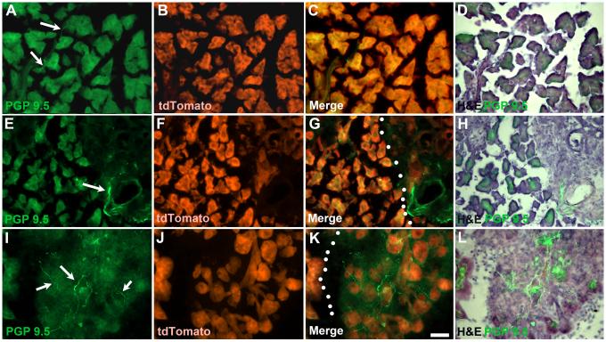
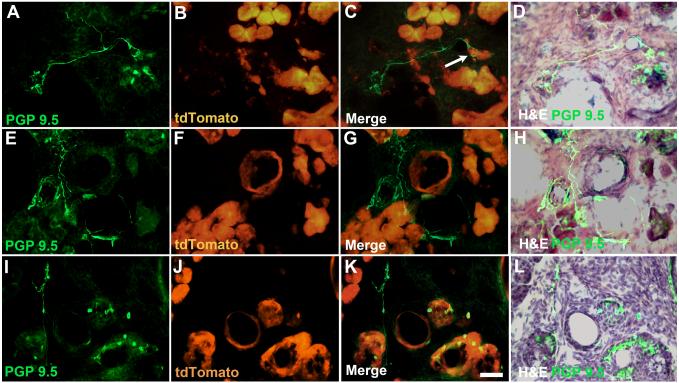
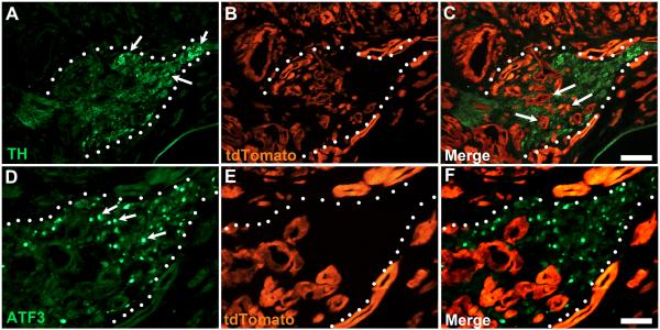
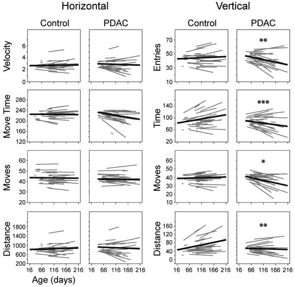
Perineural tumor invasion of intrapancreatic nerves, neurogenic inflammation, and tumor metastases along extrapancreatic nerves are key features of pancreatic malignancies. Animal studies show that chronic pancreatic inflammation produces hypertrophy and hypersensitivity of pancreatic afferents and that sensory fibers may themselves drive inflammation via neurogenic mechanisms. Although genetic mutations are required for cancer development, inflammation has been shown to be a precipitating event that can accelerate the transition of precancerous lesions to cancer. These observations led us to hypothesize that inflammation that accompanies early phases of pancreatic ductal adenocarcinoma (PDAC) would produce pathologic changes in pancreatic neurons and innervation. Using a lineage-labeled genetically engineered mouse model of PDAC, we found that pancreatic neurotrophic factor mRNA expression and sensory innervation increased dramatically when only pancreatic intraepithelial neoplasia were apparent. These changes correlated with pain-related decreases in exploratory behavior and increased expression of nociceptive genes in sensory ganglia. At later stages, cells of pancreatic origin could be found in the celiac and sensory ganglia along with metastases to the spinal cord. These results demonstrate that the nervous system participates in all stages of PDAC, including those that precede the appearance of cancer.


Figures

Figure 1
Figure 1
Distribution of PGP 9.5–positive fibers during development of PDAC. A-D) In control mice, only occasional thin PGP 9.5-positive fibers are observed (arrows) within the parenchyma of the pancreas (as indicated by expression tdTomato). E-H) By 6-8 weeks, fibrosis begins to develop and numerous PGP-positive fibers can be seen associated with blood vessels and dilated ducts (arrow). Dotted lines indicate border between fibrotic region and normal appearing pancreas (G). I-L) Significant areas of fibrosis containing dense innervation by PGP-positive fibers are present in the pancreas of 10-12 week old tPDAC mice. Dotted lines in K indicate region with developing fibrosis with atrophied acinar tissue identified based on tdTomato expression. Calibration bar = 50μm.
Figure 2
Figure 2
Examples of abnormal innervation in tPDAC mice with identifiable tumors. Hypertrophied nerves with multiple fibers were present, primarily in regions that had developed fibrosis as indicated by overlap of PGP 9.5 and loss of tdTomato-expressing cells. A-D) In some cases fiber bundles can be seen extending from atrophied/aggregated islets, E-H) near ducts or vasculature, I-L) and adjacent to areas of PanIN lesions or tumor cells. Arrow in (C) shows nerve fibers appearing to engulf tdTomato-positive cells. Calibraion bar = 50μm
Figure 3
Figure 3
PDAC induces pathological changes in celiac ganglion (indicated by dotted line). A) Celiac ganglion neurons stained with TH antibody (arrows). B) tdTomato staining reveals extensive tumor growth around and within (arrows in C) the borders of the celiac ganglion. D-F) Same celiac ganglion in A-C stained for expression of ATF3, a marker of neuronal injury. Numerous ATF3-positive neuronal nuclei are seen throughout the ganglion (arrows in A). Calibration bars A-C = 100μm; D-F = 50μm.
Figure 4
Figure 4
Pancreatic tumor surrounding the spinal cord at vertebral levels that give rise to sensory innervation of the pancreas. A) Right and left arrows indicate T13 and T9 rib, respectively, demarcating the vertebral level of the sensory ganglia giving rise to the majority of sensory innervation to the pancreas. B) Tumor, indicated by tdTomato expression, surrounds both dorsal (DR) and ventral (VR) roots of the right T11 DRG. No tdTomato-positive cells are seen in this DRG. C) Left T11 DRG containing numerous tdTomato-positive cells, presumably representing migrating tumor cells associated with the tumor formation. tdTomato-positive cells were only seen in T10 and T11 DRG on the left side. D) Left T10 DRG stained with PGP 9.5. E) Merged photomicrograph showing numerous tdTomato-positive cells interspersed between PGP 9.5-positive DRG neurons. Insert shows tdTomato–expressing cell appearing to migrating between two DRG neurons. Calibration bars A = 2mm; B&C = 100μm; C&D = 50μm.
Figure 5
Figure 5
Decreased open-field exploratory behavior in PDAC mice with increasing age. PDAC and control mice were photoelectrically monitored in an open-field arena and both horizontal and vertical movement measurements were recorded. Animals were monitored for a total of 15 minutes and data were divided into 3 blocks of 5 minutes each for analysis; data from the second block are presented as representative. * p<0.05, **p<0.01, ***p<0.001
Figure 6
Figure 6
Expression of neurotrophic factors and neurotrophic factor receptors in the pancreas of PDAC mice was measured at 3-4, 6-8, 10-12, and >16 weeks of age, normalized using Rpl13a as a reference standard, and presented as the percent expression of age-matched controls. Control n= 4 (3-4 weeks), n= 4 (6-8), n=5 (10-12), and n=6 (>16). PDAC n= 6 (3-4), n=7 (6-8), n=7 (10-12), and n=10 (>16). *p<0.05; **p<0.01; ***p<0.001. Data from >16 weeks old mice are also presented as the percent expression of controls normalized to the amount of cDNA amplified (>16Φ) and using this method of comparison, expression all neurotrophic factors and neurotrophic factor receptors except Gfrα3 is significantly increased, p<0.01.

References

    1. Ceyhan GO, Bergmann F, Kadihasanoglu M, Altintas B, Demir IE, Hinz U, et al. Pancreatic neuropathy and neuropathic pain--a comprehensive pathomorphological study of 546 cases. Gastroenterology. 2009;136:177–186.e1. - PubMed
    1. Ceyhan GO, Schäfer K-H, Kerscher AG, Rauch U, Demir IE, Kadihasanoglu M, et al. Nerve growth factor and artemin are paracrine mediators of pancreatic neuropathy in pancreatic adenocarcinoma. Ann. Surg. 2010;251:923–31. - PubMed
    1. Ceyhan GO, Giese N a, Erkan M, Kerscher AG, Wente MN, Giese T, et al. The neurotrophic factor artemin promotes pancreatic cancer invasion. Ann. Surg. 2006;244:274–81. - PMC - PubMed
    1. Ma J, Jiang Y, Jiang Y, Sun Y, Zhao X. Expression of nerve growth factor and tyrosine kinase receptor A and correlation with perineural invasion in pancreatic cancer. J. Gastroenterol. Hepatol. 2008;23:1852–9. - PubMed
    1. Zeng Q, Cheng Y, Zhu Q, Yu Z, Wu X, Huang K, et al. The Relationship between Over-expression of Glial Cell-derived Neurotrophic Factor and Its RET Receptor with Progression and Prognosis of Human Pancreatic Cancer. J. Int. Med. Res. 2008;36:656–64. - PubMed

Publication types

MeSH terms

Substances