. 2014 Mar 4;110(5):1211-20.
doi: 10.1038/bjc.2013.822. Epub 2014 Jan 21.

G-CSF and G-CSFR are highly expressed in human gastric and colon cancers and promote carcinoma cell proliferation and migration

Affiliations

G-CSF and G-CSFR are highly expressed in human gastric and colon cancers and promote carcinoma cell proliferation and migration

K T Morris et al. Br J Cancer. .

Abstract

Background: Granulocyte colony-stimulating factor (G-CSF) is a pro-inflammatory cytokine that stimulates myeloid stem cell maturation, proliferation, and migration into circulation. Despite being a known growth factor, the impact of G-CSF on solid tumours has not been well examined. G-CSF receptor (G-CSFR) is expressed by some tumours, and thus the aim of this study was to examine the expression and impact of G-CSF and G-CSFR on gastrointestinal tumours.

Methods: In this study, G-CSF expression was examined in human gastric and colon tumours and by tumour-derived stromal myofibroblasts and carcinoma cells. G-CSFR expression was examined on carcinoma cells isolated from human tissues. The effects of G-CSF on gastric and colon carcinoma cell proliferation, migration, and signalling were examined.

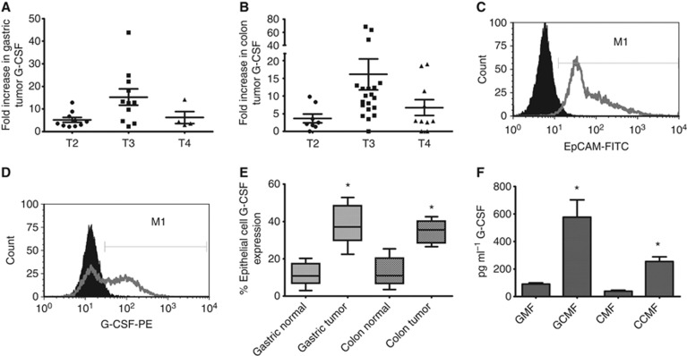
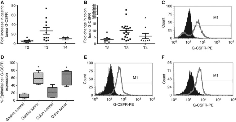
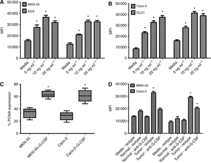
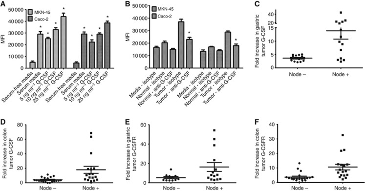
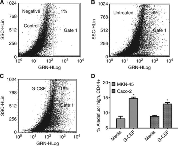
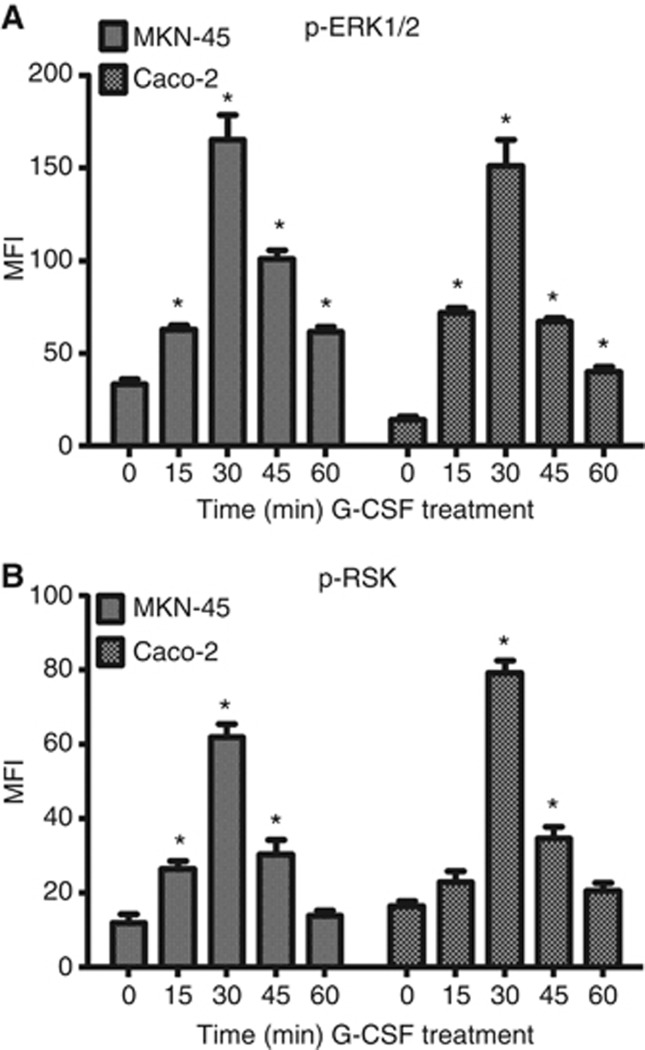
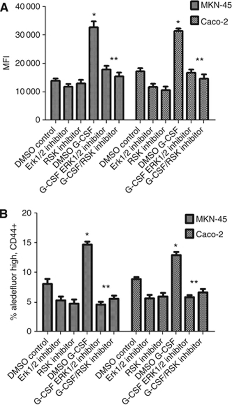
Results: G-CSFR was highly expressed in 90% of human gastric and colon carcinomas. G-CSF was also found to be highly produced by stromal myofibroblasts and carcinoma cells. Exposure of carcinoma cells to G-CSF led to increased proliferation and migration, and expansion of a sub-population of carcinoma cells expressing stem-like markers. These processes were dependent on ERK1/2 and RSK1 phosphorylation.

Conclusions: These data suggest that the G-CSF/R axis promotes gastric and colorectal cancer development and suggest they are potential tumour targets.


Figures

Figure 1
Figure 1
G-CSF is highly expressed in human tumours and by GI epithelial cells. G-CSF mRNA levels are increased in (A) gastric and (B) colon tumours compared with normal tissues as shown by tumour stage by quantitative RT–PCR. (C) Epithelial cells isolated from human tumours express EpCAM and (D) and G-CSF in representative figures. (E) In compiled data from multiple experiments, G-CSF expression is increased in epithelial cells isolated from gastric and colon tumours compared with epithelial cells from matched normal tissues when analysed by flow cytometry. G-CSF is also produced in increased amounts by tumour-derived gastric and colon cancer fibroblasts/myofibroblasts as compared with matched normal tissue-derived fibroblasts/myofibroblasts as shown in the media from such cultured cells by (F) Luminex bead array. N=8 for E and F and the mean±s.e. values are shown as the results of multiple experiments. *P<0.05.
Figure 2
Figure 2
G-CSFR is highly expressed in human tumours and by GI epithelial cells. G-CSFR mRNA levels are increased in (A) gastric and (B) colon tumours compared with normal tissues as shown by tumour stage by quantitative RT–PCR. (C) G-CSFR is expressed on epithelial cells isolated from a tumour sample and (D) expression is increased in epithelial cells isolated from gastric and colon tumours compared with epithelial cells from matched normal tissues when analysed by flow cytometry. G-CSFR is expressed on the surface of (E) MKN-45 and (F) Caco-2 cells in representative histograms compared with solid peak isotype controls. N=8 for D and the mean±s.e. values are shown as the results of multiple experiments, *P<0.05.
Figure 3
Figure 3
G-CSF induces proliferation of gastric and colon carcinoma cells. G-CSF treatment induces proliferation by CyQuant assay for DNA content of (A) MKN-45 and AGS gastric carcinoma cells, (B) Caco-2 and DLD1 colon carcinoma cells. Proliferation was further verified by (C) PCNA staining by flow cytometry. (D) Tumour-derived GMF and CMF supernatants induced proliferation of MKN-45 and Caco-2, which was decreased on adding anti-G-CSF neutralising antibodies. The mean±s.e. values are shown as the results of multiple experiments, N=8 *P<0.05 compared with untreated.
Figure 4
Figure 4
G-CSF induces migration of gastric and colon carcinoma cells. Fluorescently labelled MKN-45 and Caco-2 cells were added to the top of Fluorblock plates with 8 μm pores with (A) recombinant G-CSF and (B) normal and tumour MF supernatants with G-CSF neutralisation by monoclonal antibodies. Migration was assessed by mean fluorescence intensity. The mean±s.e. values are shown as the results of multiple experiments, N=8 *P<0.05 compared with serum-free media. Human tumour tissues from individuals that had cells migrate to lympn nodes have higher (C) G-CSF in gastric cancer, (D) G-CSF in colon cancer, (E) G-CSFR in gastric cancer, and (F) G-CSFR in colon cancer.
Figure 5
Figure 5
G-CSF increases a population of cells expressing stem-like markers in MKN-45 and Caco-2 cells. Aldefluor staining of MKN-45 cells gated on the CD44 positive population in representative dot plots as shown with (A) DEAB aldehyde dehydrogenase negative control inhibitor, (B) untreated cells, (C) G-CSF-treated cells, and (D) compiled data for MKN-45 and Caco-2 cells. For D, the mean±s.e. values are shown as the results of multiple experiments. N=8, *P<0.05.
Figure 6
Figure 6
G-CSF induces ERK1/2 and RSK signalling. Recombinant G-CSF treatment induces (A) ERK1/2 and (B) RSK1 phosphorylation. The mean±s.e. values are shown as the results of multiple experiments. N=8, *P<0.05.
Figure 7
Figure 7
Inhibition of ERK1/2 and RSK pathways reduces G-CSF-induced proliferation and the increased population expressing stem-like markers. (A) G-CSF induced cell proliferation is inhibited by ERK1/2 and RSK inhibitors as shown by fluorescent cell proliferation assay for DNA content (CyQuant) and (B) G-CSF-induced expansion populations expressing CD44 and aldehyde dehydrogenase is inhibited by ERK1/2 and RSK inhibitors. The mean±s.e. values are shown as the results of multiple experiments. N=8, *P<0.05 with G-CSF treated compared to control and **P<0.05 with inhibitors compared to G-CSF treated.

References

    1. Amariglio N, Jacob-Hirsch J, Shimoni A, Leiba M, Rechavi G, Nagler A. Changes in gene expression pattern following granulocyte colony-stimulating factor administration to normal stem cell sibling donors. Acta Haematol. 2007;117 (2:68–73. - PubMed
    1. Balkwill F, Mantovani A. Inflammation and cancer: back to Virchow. Lancet. 2001;357 (9255:539–545. - PubMed
    1. Carpentino JE, Hynes MJ, Appelman HD, Zheng T, Steindler DA, Scott EW, Huang EH. Aldehyde dehydrogenase-expressing colon stem cells contribute to tumorigenesis in the transition from colitis to cancer. Cancer Res. 2009;69 (20:8208–8215. - PMC - PubMed
    1. Dalerba P, Cho RW, Clarke MF. Cancer stem cells: models and concepts. Annu Rev Med. 2007;58:267–284. - PubMed
    1. Deng S, Yang X, Lassus H, Liang S, Kaur S, Ye Q, Li C, Wang LP, Roby KF, Orsulic S, Connolly DC, Zhang Y, Montone K, Butzow R, Coukos G, Zhang L. Distinct expression levels and patterns of stem cell marker, aldehyde dehydrogenase isoform 1 (ALDH1), in human epithelial cancers. PLoS One. 2010;5 (4:e10277. - PMC - PubMed

Publication types

MeSH terms

Substances