. 2014 Mar 14;289(11):7671-81.
doi: 10.1074/jbc.M113.545137. Epub 2014 Jan 21.

Cigarette smoke-induced Ca2+ release leads to cystic fibrosis transmembrane conductance regulator (CFTR) dysfunction

Affiliations

Cigarette smoke-induced Ca2+ release leads to cystic fibrosis transmembrane conductance regulator (CFTR) dysfunction

Julia E Rasmussen et al. J Biol Chem. .

Abstract

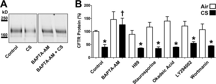
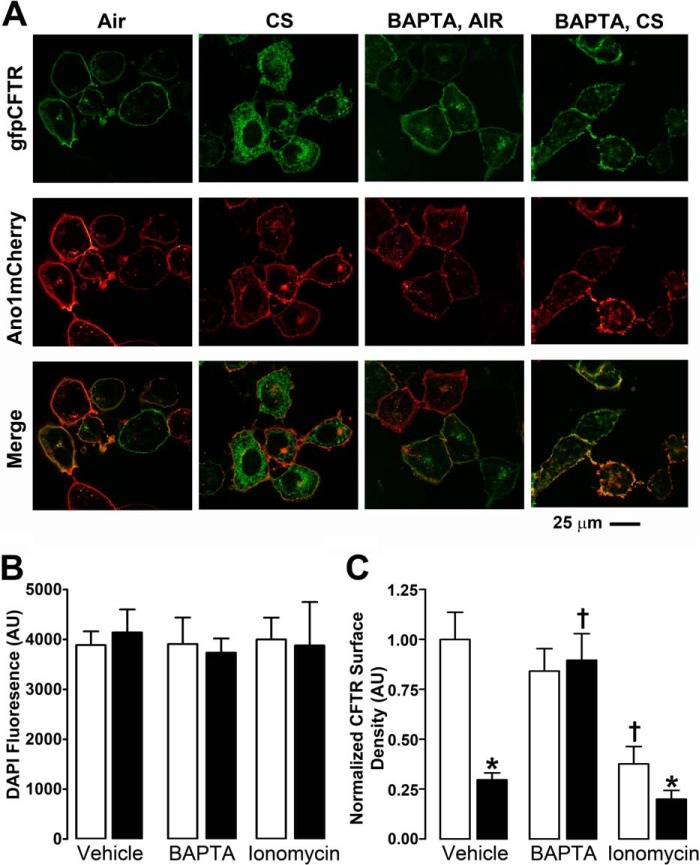
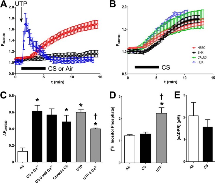
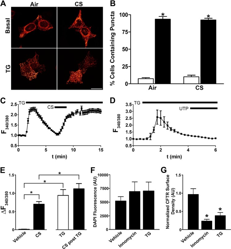
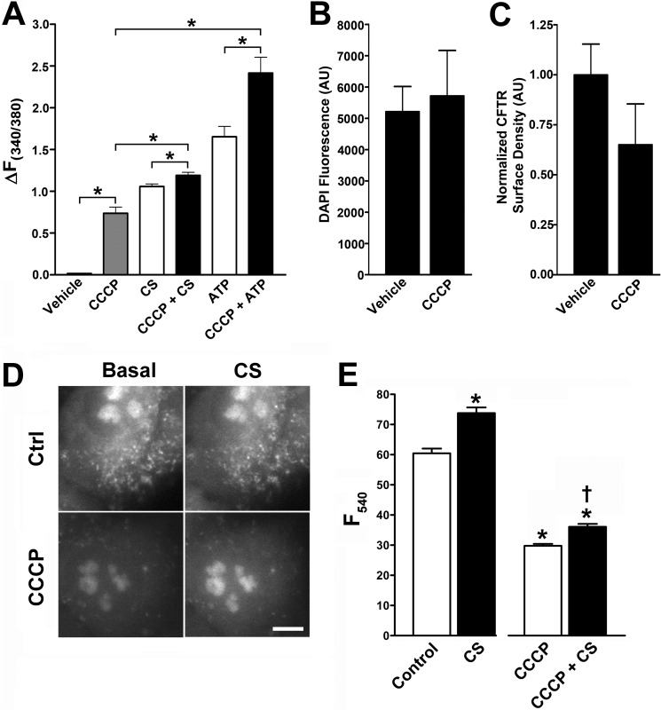
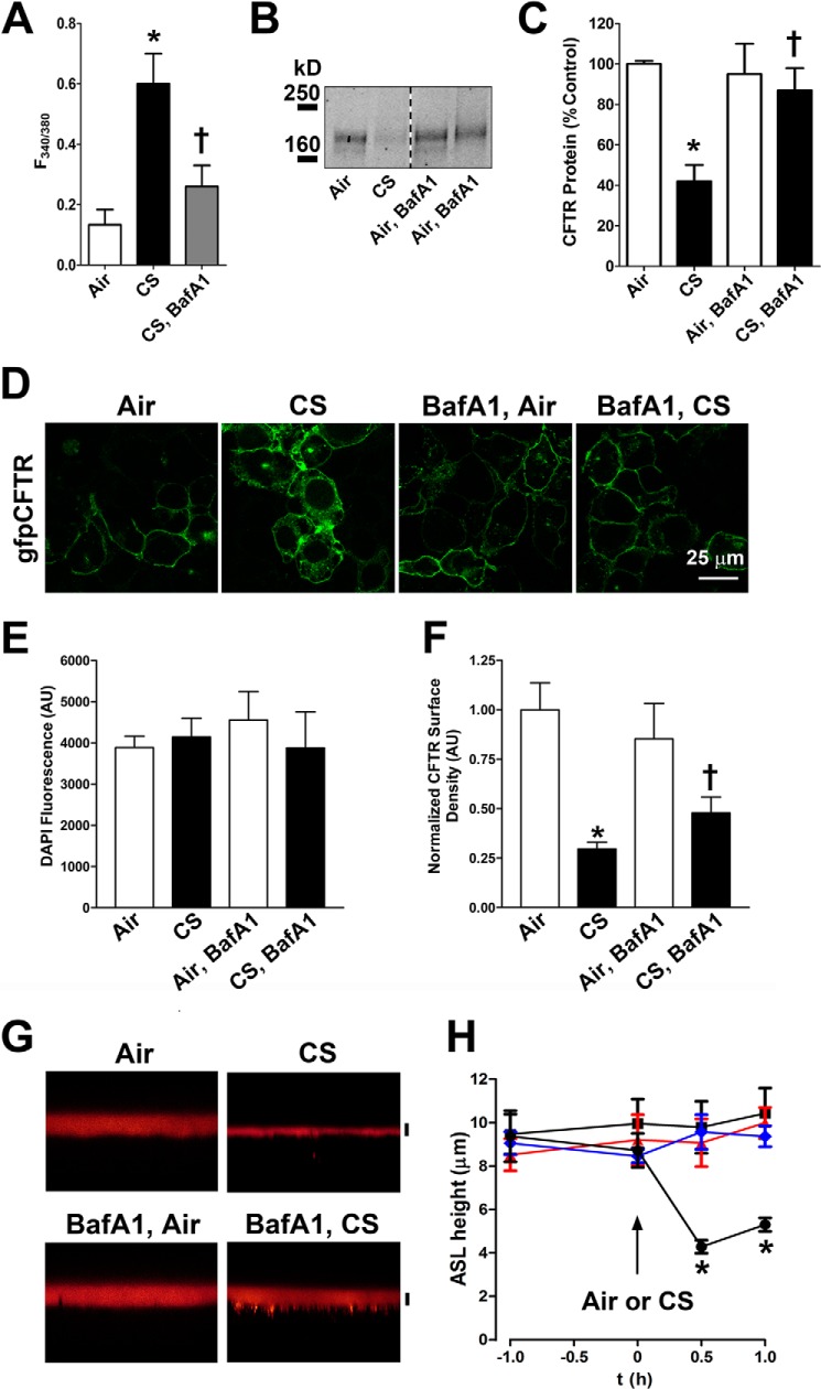
Chronic obstructive pulmonary disease affects 64 million people and is currently the fourth leading cause of death worldwide. Chronic obstructive pulmonary disease includes both emphysema and chronic bronchitis, and in the case of chronic bronchitis represents an inflammatory response of the airways that is associated with mucus hypersecretion and obstruction of small airways. Recently, it has emerged that exposure to cigarette smoke (CS) leads to an inhibition of the cystic fibrosis transmembrane conductance regulator (CFTR) Cl(-) channel, causing airway surface liquid dehydration, which may play a role in the development of chronic bronchitis. CS rapidly clears CFTR from the plasma membrane and causes it to be deposited into aggresome-like compartments. However, little is known about the mechanism(s) responsible for the internalization of CFTR following CS exposure. Our studies revealed that CS triggered a rise in cytoplasmic Ca(2+) that may have emanated from lysosomes. Furthermore, chelation of cytoplasmic Ca(2+), but not inhibition of protein kinases/phosphatases, prevented CS-induced CFTR internalization. The macrolide antibiotic bafilomycin A1 inhibited CS-induced Ca(2+) release and prevented CFTR clearance from the plasma membrane, further linking cytoplasmic Ca(2+) and CFTR internalization. We hypothesize that CS-induced Ca(2+) release prevents normal sorting/degradation of CFTR and causes internalized CFTR to reroute to aggresomes. Our data provide mechanistic insight into the potentially deleterious effects of CS on airway epithelia and outline a hitherto unrecognized signaling event triggered by CS that may affect the long term transition of the lung into a hyper-inflammatory/dehydrated environment.

Keywords: Chronic Obstructive Pulmonary Disease (COPD); Cystic Fibrosis; Epithelium; Ion Channels; Lysosomes.


Figures

FIGURE 1.
FIGURE 1.
Screening for antagonists of CS-induced CFTR diminution by Western blot. BHKCFTR cultures were exposed to 10 air or CS puffs. A, representative Western blot for mature CFTR following CS exposure ± pretreatment with 5 μm BAPTA-AM. B, mean densitometry of CFTR following pretreatment with either BAPTA-AM or a variety of inhibitors followed by CS exposure. All n = 3. Open bars, air exposure. Closed bars, CS exposure. * denotes p < 0.05 different from control/air. † denotes p < 0.05 different from control/CS.
FIGURE 2.
FIGURE 2.
CS exposure leads to CFTR internalization that is sensitive to Ca2+ chelation. BHKCFTR cultures were exposed to 10 air or CS puffs. A, representative confocal micrographs from three individual experiments showing that gfpCFTR (green) but not Ano1mCherry (red) is internalized by CS exposure and that this response is abrogated following Ca2+ chelation with BAPTA-AM. B, CS exposure does not affect DAPI fluorescence, an indicator of total BHKCFTR cell number. C, surface CFTR fluorescence from BHKCFTR cells (normalized to DAPI and vehicle control) following air or CS exposure in the presence and absence of BAPTA or ionomycin. n = 18–32 cultures from four separate experiments. Open bars, air exposure. Closed bars, CS exposure. * denotes p < 0.05 different from control/air. † denotes p < 0.05 different from control/CS. AU, arbitrary units.
FIGURE 3.
FIGURE 3.
Cigarette smoke exposure increases cytosolic Ca2+ levels. HBECs were preloaded with Fura2 and then exposed to 10 puffs of CS or air in situ under thin film conditions in an environmental chamber on an epifluorescent microscope. A, mean change in Fura2 ratio (F340/380) as an indicator of cytosolic Ca2+ with time before/after 10 puffs of air (○) or smoke (□). Exposure occurred during the period indicated by the horizontal bar, and afterward, CS was flushed out of the chamber and exchanged for room air. Both n = 12. As an additional control, UTP was added to increase Ca2+ (Δ; n = 6). B, cytosolic Ca2+ was elevated following CS exposure in a variety of cell types. Mean change in Fura2 ratio (F340/380) is shown over time as an indicator of cytosolic Ca2+. ●, HBECs; ■, BHKCFTR cells; ▴, CALU3 cells; ▾, HEK293T cells. C, bar graph showing mean change in F340/380 after 10 min of air (open bars), CS, removal/chelation of extracellular Ca2+ followed by CS, chronic CS exposure (closed bars), and UTP ± extracellular Ca2+ (gray bars). All n ≥6. D, [3H]inositol phosphate accumulation in HBECs after air, CS, or UTP exposure. All n = 4. E, cyclic ADP-ribose (cADPR) levels in CALU3 cells before/after CS exposure. All n = 4. *, p < 0.05 different from air control. † denotes p < 0.05 different from UTP.
FIGURE 4.
FIGURE 4.
CS exposure does not elicit Ca2+ release from the endoplasmic reticulum. A, confocal micrographs of STIM1mCherry expressed in HEK293T cells and imaged under basal conditions, following 1 μm thapsigargin (TG) addition or after 10 puffs of CS ± thapsigargin. B, percentage of HEK293T cells containing puncta. C and D, absolute F340/380 levels in Fura2-loaded HEK293T cells sequentially exposed to 1 μm thapsigargin followed by 10 puffs of CS or 10 μm UTP addition, respectively (both n = 8). E, change in Fura2 emission ratio (ΔF340/380) in HEK293T cells exposed to vehicle, 10 puffs of CS, 1 μm thapsigargin, or thapsigargin followed by CS (all n = 8). F, ionomycin and thapsigargin do not affect DAPI fluorescence, an indicator of total HEK293T cell number. G, surface CFTR expression in HEK293T cells (normalized to DAPI and vehicle control) following ionomycin or thapsigargin treatment. Vehicle, n = 26; ionomycin, n = 11; thapsigargin, n = 10. * denotes significance (p < 0.05) across groups. AU, arbitrary units.
FIGURE 5.
FIGURE 5.
CS exposure does not elicit Ca2+ release from mitochondrial stores. A, bar graph showing change in Fura2 fluorescence (ΔF340/380) in HEK293T cells following vehicle, CCCP alone (gray bars), 10 puffs of CS, or 10 μm ATP (open bars) ± 1 h of pretreatment with 10 μm CCCP (closed bars). Vehicle, n = 30; CS, n = 43; CCCP + CS = 45; ATP, n = 18; CCCP + ATP, n = 15. *, p < 0.05. B, CCCP Fura-2 does not affect DAPI fluorescence, an indicator of total HEK293T cell number. C, CFTR surface expression is not affected by CCCP. All, n = 10. AU, arbitrary units. D, images of Rhod-2-loaded HEK293T cells ± CCCP and CS. Ctrl, control. E, bar graph showing mean Rhod-2 emission in mitochondria from HEK293T cells exposed to CCCP followed by 10 puffs of CS, respectively (CT, n = 69; CCCP, n = 67). Open bars, control. Closed bars, CS or ATP. *, p < 0.05 different from control. † denotes p < 0.05 different from CCCP.
FIGURE 6.
FIGURE 6.
Peri-lysosomal Ca2+ levels increase with CS exposure. HEK293T cells expressing LAMP1-YCam3.6 were exposed to 10 puffs of air or CS and then fixed immediately after exposure in 4% PFA and prepared for imaging. A, images of CFP (pseudocolored gray) and YFP (pseudocolored red) before and after photobleaching of YFP. B, resultant FRET image taken from A. C, mean change in FRET efficiency (%E) following CS (closed bars) or air (open bars). As controls, CFP linked to YFP by five glycines (CFP-5G-YFP) and unlinked CFP/YFP were also employed. Numbers of cells imaged are in parentheses. *, p < 0.05 different from air.
FIGURE 7.
FIGURE 7.
Bafilomycin-A1 pretreatment abolishes CS-induced increases in cytosolic Ca2+ and reduces diminution/internalization of CFTR. A, bar graph showing mean changes in cytosolic Ca2+ (F340/380) in HBECs following air (open bars; n = 12), CS (closed bars; n = 12), or bafilomycin pretreatment (400 nm) followed by CS (gray bars; n = 12). B, Western blot showing CFTR ± CS in the presence/absence of 400 nm bafilomycin in BHKCFTR cells. C, mean densitometry taken from B. All n = 3. D, confocal micrographs showing gfpCFTR (green) following air and CS exposure ± bafilomycin. E, CS and bafilomycin exposure do not affect DAPI fluorescence, an indicator of total BHKCFTR cell number. AU, arbitrary units. F, surface CFTR fluorescence from BHKCFTR cells (normalized to DAPI and vehicle control) following air or CS exposure in the presence and absence of 300 nm bafilomycin. n = 12–24 cultures from three separate experiments. G, typical confocal micrographs showing ASL height after air or CS exposure ± bafilomycin pretreatment (300 nm/30 min). H, graph taken from G showing mean changes in ASL height with time before and after air or CS exposure. ■, air, vehicle; ▴, air, BAF; ●, CS, vehicle; ♦, CS, BAF. All n = 8. * denotes p < 0.05 different from control (air). † denotes p < 0.05 different from CS.

Similar articles

Cited by

References

    1. Vareille M., Kieninger E., Edwards M. R., Regamey N. (2011) The airway epithelium: soldier in the fight against respiratory viruses. Clin. Microbiol. Rev. 24, 210–229 - PMC - PubMed
    1. Bartlett J. A., Fischer A. J., McCray P. B., Jr. (2008) Innate immune functions of the airway epithelium. Contrib. Microbiol. 15, 147–163 - PubMed
    1. Schmid A., Clunes L. A., Salathe M., Verdugo P., Dietl P., Davis C. W., Tarran R. (2011) Nucleotide-mediated airway clearance. Subcell. Biochem. 55, 95–138 - PubMed
    1. Com G., Clancy J. P. (2009) Adenosine receptors, cystic fibrosis, and airway hydration. Handb. Exp. Pharmacol. 363–381 - PubMed
    1. Chmiel J. F., Davis P. B. (2003) State of the art: why do the lungs of patients with cystic fibrosis become infected and why can't they clear the infection? Respir. Res. 4, 8. - PMC - PubMed

Publication types

MeSH terms

LinkOut - more resources