. 2014 May;32(5):1289-300.
doi: 10.1002/stem.1633.

Alk2 regulates early chondrogenic fate in fibrodysplasia ossificans progressiva heterotopic endochondral ossification

Affiliations

Alk2 regulates early chondrogenic fate in fibrodysplasia ossificans progressiva heterotopic endochondral ossification

Andria L Culbert et al. Stem Cells. 2014 May.

Abstract

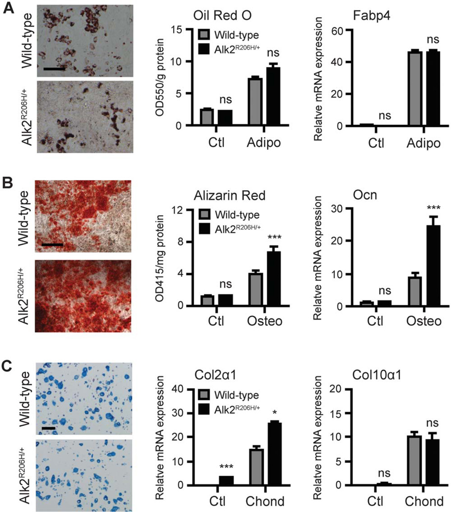
Bone morphogenetic protein (BMP) signaling is a critical regulator of cartilage differentiation and endochondral ossification. Gain-of-function mutations in ALK2, a type I BMP receptor, cause the debilitating disorder fibrodysplasia ossificans progressiva (FOP) and result in progressive heterotopic (extraskeletal) endochondral ossification within soft connective tissues. Here, we used murine mesenchymal progenitor cells to investigate the contribution of Alk2 during chondrogenic differentiation and heterotopic endochondral ossification (HEO). Alk2(R206H/+) (gain-of-function), Alk2(CKO) (loss-of-function), and wild-type mouse embryonic fibroblasts were evaluated for chondrogenic potential. Chondrogenic differentiation was accelerated in Alk2(R206H/+) cells, due in part to enhanced sensitivity to BMP ligand. In vivo, Alk2(R206H/+) cells initiated robust HEO and recruited wild-type cell contribution. Despite expression of other type I BMP receptors (Alk3 and Alk6), chondrogenesis of Alk2(CKO) cells was severely impaired by absence of Alk2 during early differentiation. Alk2 is therefore a direct regulator of cartilage formation and mediates chondrogenic commitment of progenitor cells. These data establish that at least one effect of ALK2 gain-of-function mutations in FOP patients is enhanced chondrogenic differentiation which supports formation of heterotopic endochondral bone. This establishes ALK2 as a plausible therapeutic target during early chondrogenic stages of lesion formation for preventing heterotopic bone formation in FOP and other conditions.

Keywords: Alk2; Bone morphogenetic protein signaling; Chondrogenesis; Endochondral ossification; Fibrodysplasia ossificans progressiva.


Conflict of interest statement

Disclosure of Potential Conflicts of Interest

The authors indicate no potential conflicts of interest.

Figures

Figure 1
Figure 1
Enhanced BMP signaling of Alk2R206H/+ mouse embryonic fibroblasts (MEFs). (A): Expression of type I and type II BMP receptor mRNAs in undifferentiated MEFs was quantified and normalized to Gapdh levels. (B): Immunoblot detection of pSmad1/5/8 in the absence and presence of BMP4. Quantified pSmad1/5/8 was normalized to β-actin. Lanes were run on the same gel but are noncontiguous. (C): BMP target genes expression without BMP (left) and in response to BMP4 (15 ng/ml) for 2 hours (right) detected by qRT-PCR and normalized to Gapdh levels. (D): Growth curves of wild-type and Alk2R206H/+ MEFs. All data represent mean ± SEM; **, p ≤ .01; ***, p ≤ .001; ns = not significant. Abbreviation: BMP, bone morphogenetic protein.
Figure 2
Figure 2
Mesenchymal multipotency of mouse embryonic fibroblasts (MEFs). (A): MEFs in adipogenic media (14 days) were stained with oil red O to visualize lipid droplets and quantified. Fabp4 expression was normalized to Gapdh. (B): MEFs in osteogenic media (14 days) were stained with Alizarin Red to visualize calcium deposition and quantified. Ocn expression was normalized to 18S rRNA. (C): MEFs in chondrogenic media containing BMP4 (100 ng/ml) (14 days) were stained with Alcian blue to visualize chondrocyte morphology and matrix. Col2α1 and Col10α1 expressions were normalized to 18s rRNA. All data represent mean ± SEM; n = 3; *, p ≤ .05; ***, p ≤ .001; ns = not significant. Scale bars = 100 µm.
Figure 3
Figure 3
Alk2R206H/+ does not predispose cells toward chondrogenesis. (A): Expression of Prrx1 and Fsp1 (fibroblast) and Nxk3.2 and Sox5/6/9 (prechondrogenic) mRNAs were quantified in undifferentiated mouse embryonic fibroblasts (MEFs) and normalized to 18S rRNA. (B): MEFs cultured in three-dimensional alginate spheres with chondrogenic media in the absence (control) or presence of BMP4 (100 ng/ml) for 21 days were sectioned and stained with Alcian blue to visualize chondrocyte morphology and matrix. All data represent mean ± SEM; ns = not significant.
Figure 4
Figure 4
Increased sensitivity and accelerated chondrogenic differentiation by Alk2R206H/+. (A): Mouse embryonic fibroblasts (MEFs) in three-dimensional chondrogenic culture with increasing BMP4 concentrations were stained with Alcian blue (7 days). (B): Alcian bluepositive cells were quantified as the percentage of chondrocytes relative to total cells in field. (C): MEFs in chondrogenic media with 15 ng/ml BMP4 were detected for collagen type II over 14 days. Negative control is without primary antibody. (D): Expression of chondrocyte-specific genes over time was quantified by qRT-PCR and normalized to 18S rRNA. All data represent mean ± SEM; *, p ≤ .05; **, p ≤ .01; ***, p ≤ .001; ns = not significant. Scale bars = 100 µm.
Figure 5
Figure 5
Alk2R206H/+ cells induce heterotopic endochondral ossification (HEO) in vivo. (A): Serial sections through implants were examined for endochondral ossification. Hematoxylin and eosin staining shows implant (im) adjacent to normal skeletal muscle (sm) tissue. Safranin-O (red) detects chondrocytes. Red fluorescent Qdots, from labeled donor cells, are present within chondrocytes of Alk2R206H/+ cell implants (arrows). GFP antibody detection identifies infiltrated GFP-tagged host cells within the implants (all conditions) and a mixture of GFP (donor) and GFP+ (host) chondrocytes participating in HEO. Alcian blue/orange G staining differentiates cartilage (blue) from surrounding fragments of mature bone (bright pink; arrows). Scale bars = 100 µm. (B): Wild-type or Alk2R206H/+ mouse embryonic fibroblasts with 500ng BMP4 were injected into mouse hind limb muscles (right); the contralateral limb (left) was injected with BMP alone and microCT evaluated ossification after 21 days. Insets highlight detected mineralization in regions of cell implants. Where ectopic mineralization was detected the total mineralized volume (mm3) was quantified using Scanco software and is shown numerically above the sample. Data represent mean ± SEM; ***, p ≤ .001. Scale bars = 1 µm. Abbreviation: GFP, green fluorescent protein.
Figure 6
Figure 6
Alk2 expression is required during early chondrogenesis. (A): Expression of Alk2, Alk3, and Alk6 type I BMP receptors during early chondrogenic differentiation of wild-type mouse embryonic fibroblasts (MEFs) was determined by qRT-PCR and normalized to 18S rRNA. (B): BMP signaling in wild-type and Alk2CKO MEFs in the presence of BMP4 was detected by immunoblot analysis of pSmad1/5/8 and normalized to α-tubulin. (C): Schematic shows timing of Alk2 knockout (arrow) before or during chondrogenic differentiation. Chondrocyte-specific markers were assessed by qRT-PCR after 14 days and normalized to β-2-microglobulin mRNA. All data represent mean ± SEM; *, p ≤ .05; **, p ≤ .01; ***, p ≤ .001; ns = not significant.

References

    1. Mackie EJ, Tatarczuch L, Mirams M. The skeleton: A multi-functional complex organ: The growth plate chondrocyte and endochondral ossification. J Endocrinol. 2011;211:109–121. - PubMed
    1. Shore EM, Kaplan FS. Inherited human diseases of heterotopic bone formation. Nat Rev Rheumatol. 2010;6:518–527. - PMC - PubMed
    1. Forsberg JA, Potter BK. Heterotopic ossification in wartime wounds. J Surg Orthop Adv. 2010;19:54–61. - PubMed
    1. Cremin B, Connor JM, Beighton P. The radiological spectrum of fibrodysplasia ossificans progressiva. Clin Radiol. 1982;33:499–508. - PubMed
    1. Kaplan FS, Xu M, Seemann P, et al. Classic and atypical fibrodysplasia ossificans progressiva (Fop) phenotypes are caused by mutations in the bone morphogenetic protein (Bmp) type I receptor Acvr1. Hum Mutat. 2009;30:379–390. - PMC - PubMed

Publication types

MeSH terms

Substances