. 2014 Jan 15;28(2):127-39.
doi: 10.1101/gad.231969.113.

Mechanical motion promotes expression of Prg4 in articular cartilage via multiple CREB-dependent, fluid flow shear stress-induced signaling pathways

Affiliations

Mechanical motion promotes expression of Prg4 in articular cartilage via multiple CREB-dependent, fluid flow shear stress-induced signaling pathways

Hiroyasu Ogawa et al. Genes Dev. .

Abstract

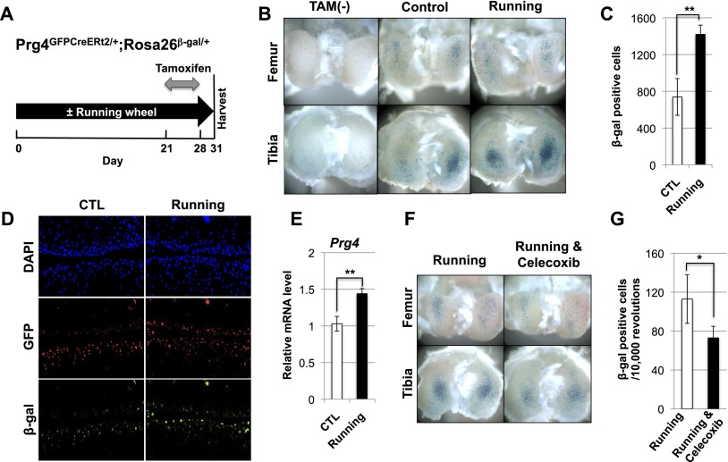
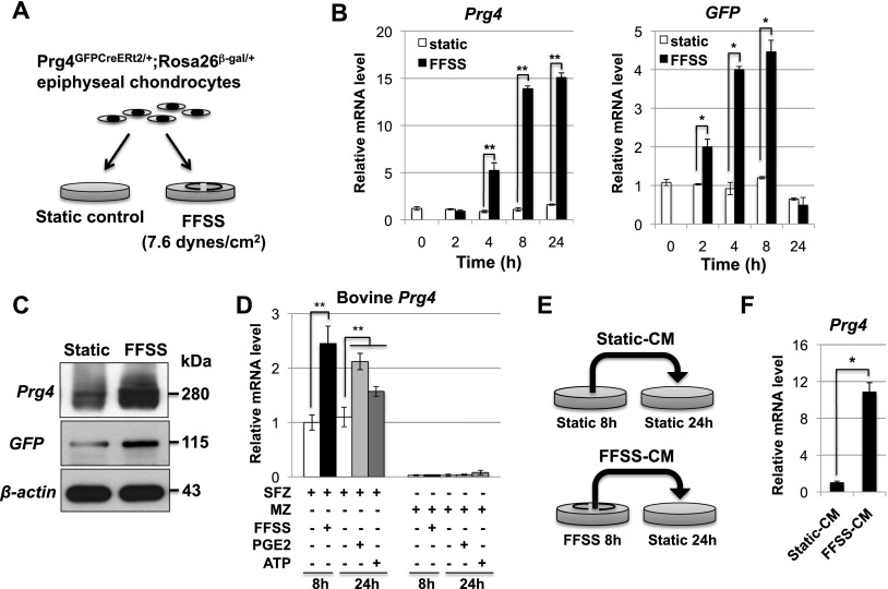
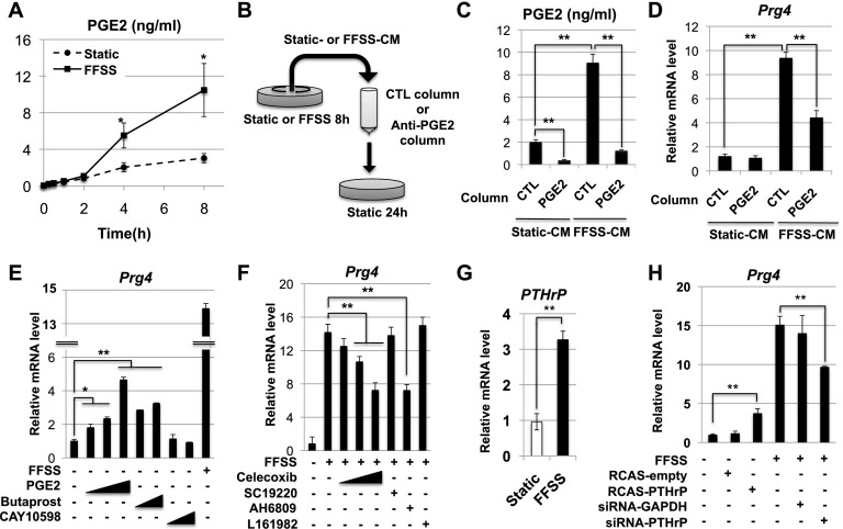
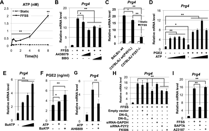
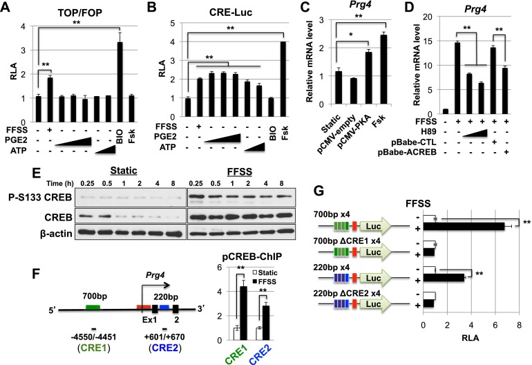
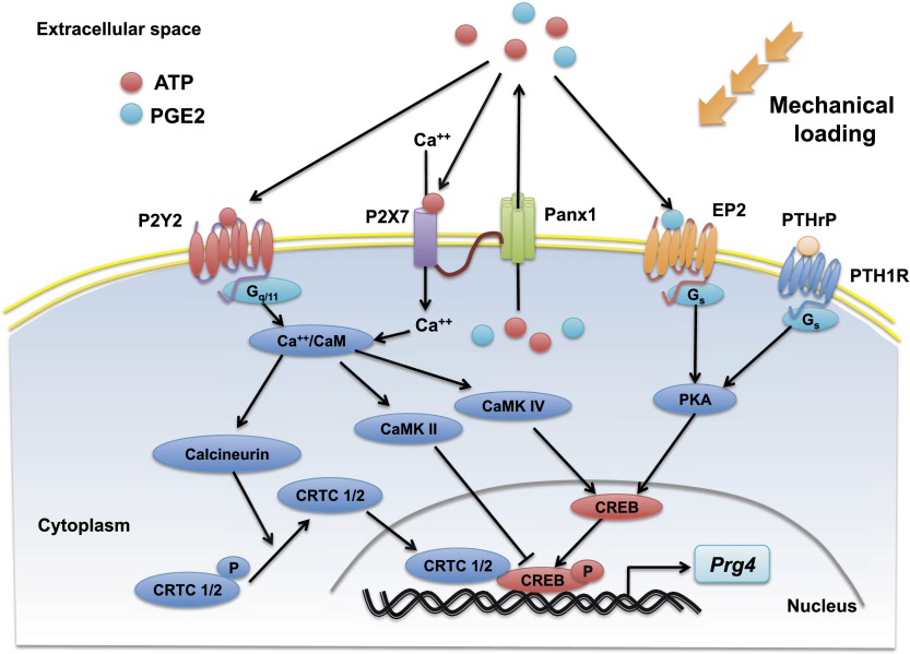
Lubricin is a secreted proteoglycan encoded by the Prg4 locus that is abundantly expressed by superficial zone articular chondrocytes and has been noted to both be sensitive to mechanical loading and protect against the development of osteoarthritis. In this study, we document that running induces maximal expression of Prg4 in the superficial zone of knee joint articular cartilage in a COX-2-dependent fashion, which correlates with augmented levels of phospho-S133 CREB and increased nuclear localization of CREB-regulated transcriptional coactivators (CRTCs) in this tissue. Furthermore, we found that fluid flow shear stress (FFSS) increases secretion of extracellular PGE2, PTHrP, and ATP (by epiphyseal chondrocytes), which together engage both PKA- and Ca(++)-regulated signaling pathways that work in combination to promote CREB-dependent induction of Prg4, specifically in superficial zone articular chondrocytes. Because running and FFSS both boost Prg4 expression in a COX-2-dependent fashion, our results suggest that mechanical motion may induce Prg4 expression in the superficial zone of articular cartilage by engaging the same signaling pathways activated in vitro by FFSS that promote CREB-dependent gene expression in this tissue.

Keywords: CREB; PGE2; PTHrP; Prg4/lubricin; articular cartilage; extracellular ATP.


Figures

Figure 1.
Figure 1.
Wheel running increases Prg4 expression. (A) Three-month-old female Prg4GFPCreERt2/+; Rosa26floxlacZ/+ mice were housed in either the absence or presence of a running wheel for 31 d. The animals were injected with tamoxifen daily from day 22 to 28 and sacrificed for X-gal staining at day 31. (B) Whole-mount X-gal staining of the knee joints taken from either corn-oil-injected (TAM−) or tamoxifen-injected Prg4GFPCreERt2/+; Rosa26floxlacZ/+ mice that were housed either without (Control) or with (Running) a running wheel. (C) Sections of the knee joints depicted in B (n = 6 running or 6 control mice). For each mouse, the total number of β-galactosidase-expressing cells in five sections of the lateral condyles and five sections of the medial condyles in both knees was quantitated. (D) The knees of either control or running Prg4GFPCreERt2/+; Rosa26floxlacZ/+ mice (treated as outlined in A) were sectioned and immunostained with both anti-GFP and anti-β-galactosidase antibodies. (E) Endogenous expression of Prg4 relative to β-actin was determined by RT-qPCR. (F) Whole-mount X-gal staining of the knee joints of running Prg4GFPCreERt2/+; Rosa26floxlacZ/+ mice administered either a control gavage solution (Running) or such a solution containing Celecoxib (Running & Celecoxib). (G) The knees of either six control or six Celecoxib-treated Prg4GFPCreERt2/+; Rosa26floxlacZ/+ mice (all housed with running wheels) were stained with X-gal and sectioned. The total number of β-galactosidase-expressing cells in five sections of the lateral condyles and five sections of the medial condyles was quantitated and normalized to the number of rotations of the running wheels (n = 6 control mice, n = 6 Celecoxib-treated mice). Throughout this and the following figures, significance was calculated using Student's t-test. (*) P < 0.05; (**) P < 0.01. Error bar indicates standard error of the mean.
Figure 2.
Figure 2.
FFSS promotes the secretion of a signaling molecule that in turn induces Prg4 expression in chondrocytes. (A) Epiphyseal chondrocytes were isolated from 5-d-old Prg4GFPCreERt2/+; Rosa26floxlacZ/+ animals and cultured in tissue culture dishes on either a static surface or a rotary shaker (which delivered ∼7.6 dyn/cm2). (B) Expression of both the Prg4WT and the Prg4GFPCreERt2 alleles in epiphyseal chondrocytes cultured under either static or FFSS conditions were assayed relative to β-actin by RT-qPCR. (C) Either conditioned medium (top panel) or total cell lysates (middle and bottom panels) were collected from epiphyseal chondrocytes cultured under either static or FFSS conditions for 24 h, and expression of the indicated proteins was assayed by Western blot. (D) Either FFSS, PGE2, or extracellular ATP was administered (for the indicated time period) to either superficial zone (SFZ) or middle zone (MZ) newborn bovine articular chondrocytes. Prg4 expression was assayed relative to that of 18S rRNA by RT-qPCR. (E,F) Medium conditioned by murine epiphyseal chondrocytes cultured under either static or FFSS conditions for 8 h was applied to new plates of chondrocytes cultured under static conditions for 24 h. Expression of endogenous Prg4 was assayed relative to β-actin by RT-qPCR. Throughout this figure: (*) P < 0.05; (**) P < 0.01. Error bar indicates standard error of the mean.
Figure 3.
Figure 3.
FFSS induces Prg4 expression via both PGE2- and PTHrP-dependent pathways. (A) Concentration of PGE2 secreted into conditioned medium harvested from epiphyseal chondrocytes cultured under either FFSS or static conditions for the indicated time periods. (B) Either control- or FFSS-conditioned medium was poured over a column containing either control IgG or anti-PGE2. Flow-through material that was not bound to the column was applied to new epiphyseal chondrocytes cultured under static conditions. (C,D) Depletion of >90% of PGE2 from FFSS-conditioned medium (C) attenuated the ability of this medium to induce the expression of Prg4 in static chondrocytes by ∼50% (D). (E) Epiphyseal chondrocytes were cultured under either FFSS or static conditions with increasing concentrations of either PGE2 (0.01, 0.1, and 1 μM), the EP2 agonist Butaprost (1 and 10 μM), or the EP4 agonist CAY10598 (1 and 10 μM). Prg4 expression was assayed relative to β-actin by RT-qPCR. (F) Epiphyseal chondrocytes were cultured under either static or FFSS conditions with increasing concentrations of either the COX-2 inhibitor Celecoxib (0.1, 1, 10 μM), the EP1 antagonist SC19220 (10 μM), the EP1/2 antagonist AH6809 (1 μM), or the EP4 antagonist L161982 (10 μM). Prg4 expression was assayed relative to β-actin by RT-qPCR. (G) Epiphyseal chondrocytes were isolated from 5-d-old mice and cultured under either FFSS or static conditions for 8 h. Gene expression was assayed relative to β-actin by RT-qPCR. (H) Epiphyseal chondrocytes were cultured under either FFSS or static conditions after transfection with either an RCAS empty expression vehicle, RCAS-encoding PTHrP, siRNA targeting GAPDH, or siRNA targeting PTHrP. Gene expression of Prg4 (relative to β-actin) was assayed by RT-qPCR. Throughout this figure: (*) P < 0.05; (**) P < 0.01. Error bar indicates standard error of the mean.
Figure 4.
Figure 4.
FFSS induces expression of Prg4 in articular chondrocytes via both ATP and PGE2 signaling pathways. (A) Concentration of ATP secreted into conditioned medium harvested from epiphyseal chondrocytes cultured under either FFSS or static conditions for the indicated time periods. (B) Administration of the P2X7 antagonists A438079 (1 and 10 μM) or Brilliant Blue-G (BBG; 1 and 10 μM) to epiphyseal chondrocytes significantly decreased induction of Prg4 by FFSS. (C) FFSS-mediated induction of Prg4 expression in epiphyseal chondrocytes isolated from either wild-type Balb/c mice (with P451 in P2X7), wild-type C57Bl/6J mice (with P451L in P2X7), or P2X7−/− mice (in a C57Bl/6J background). (D) Increasing amounts of ATP (0.01, 0.1, 1, and 10 μM) were administered to static chondrocytes in either the absence or presence of PGE2 (0.1 μM). (E) Increasing amounts (1, 10, and 100 uM) of the P2X7 agonist BzATP robustly induce the expression of Prg4 in static chondrocytes. (F) Administration of either ATP (1 mM) or BzATP (100 μM) to static chondrocytes induces secretion of PGE2 into the medium. (G) Induction of Prg4 expression in epiphyseal chondrocytes by extracellular ATP was attenuated by the EP1/2 antagonist AH6809 (1 μM). (H) FFSS-mediated induction of Prg4 in epiphyseal chondrocytes was assayed in the presence of either dominant-negative (DN) forms of either Gq(α) or G11(α), siRNAs directed against either GAPDH (control) or P2Y2, or the calcineurin antagonist FK506. (I) Addition of the Ca++ chelator BAPTA attenuated induction of Prg4 by FFSS, and, conversely, addition of the Ca++ ionophore A23187 induced the expression of Prg4 in static chondrocytes. Throughout this figure, Prg4 expression was assayed relative to β-actin by RT-qPCR. (*) P < 0.05; (**) P < 0.01. Error bar indicates standard error of the mean.
Figure 5.
Figure 5.
FFSS induces Prg4 expression via the PKA/CREB signaling pathway. (A) Epiphyseal chondrocytes were cotransfected with a luciferase reporter driven by either reiterated consensus TCF-binding sites (TOP-firefly luciferase) or mutated TCF-binding sites (FOP-firefly luciferase) plus SV40 Renilla luciferase. The chondrocytes were cultured in the presence of either FFSS, exogenous PGE2 (0.01, 0.1, and 1 μM), ATP (0.1 and 1 mM), the Wnt agonist BIO (10 μM), or forskolin (FSK; 10 μM). The ratio of TOP/FOP luciferase activity (normalized to that of SV40 Renilla luciferse) is shown for each treatment regimen. (B) Epiphyseal chondrocytes were cotransfected with a luciferase reporter driven by reiterated consensus CRE-binding sites (CRE-firefly luciferase) plus SV40 Renilla luciferase. The chondrocytes were cultured in the presence of either FFSS, exogenous PGE2 (0.01, 0.1, and 1 μM), ATP (0.1 and 1 mM), the Wnt agonist BIO (10 μM), or FSK (1 μM). Relative luciferase activity (RLA; normalized to that of SV40 Renilla luciferase) is shown for each treatment regimen. (C) Transfection of epiphyseal chondrocytes (cultured under static conditions) with an expression vehicle encoding an activated form of PKA or treatment with FSK was sufficient to induce expression of Prg4. Prg4 expression was assayed relative to β-actin by RT-qPCR. (D) Administration of the PKA antagonist H89 or transfection with an expression vehicle encoding ACREB attenuated the ability of FFSS to induce Prg4 expression in epiphyseal chondrocytes. Prg4 expression was assayed relative to β-actin by RT-qPCR. (E) FFSS increases steady levels of both total and phospho-S133 CREB in epiphyseal chondrocytes. (F) FFSS increases binding of phospho-CREB to two conserved CRE-binding sites (CRE1 and CRE2) that surround the TSS of Prg4 in epiphyseal chondrocytes, as detected by chromatin immunoprecipitation. (G) Epiphyseal chondrocytes were cotransfected with a firefly luciferase reporter driven by the Prg4 basal promoter plus four repeats of Prg4 sequences encompassing either wild-type or mutant CRE1- or CRE2-binding sites plus SV40-Renilla luciferase. The cells were cultured under either static or FFSS conditions, and RLA is displayed. In both cases, mutation of the CRE consensus core-binding site completely eliminated induction of these reporters by FFSS. Throughout this figure: (*) P < 0.05; (**) P < 0.01. Error bar indicates standard error of the mean.
Figure 6.
Figure 6.
FFSS/running both decrease phosphorylation of S-151 in CRTC1 and induce nuclear translocation of CRTC1 in articular cartilage. (A) Western analysis for either phospho-S-151 CRTC1, total CRTC1, or β-actin in mouse epiphyseal chondrocytes that had been cultured under either FFSS or static conditions. (B) Cellular localization of CRTC1 (by immunofluorescence) in epiphyseal chondrocytes cultured under either static conditions (in either the absence or presence of extracellular ATP) or FFSS conditions. Quantitation of cellular localization of CRTC1 is displayed on the right. (C) Cellular localization of phospho-S133 CREB (by immunofluorescence) in epiphyseal chondrocytes cultured under either static or FFSS conditions. Quantitation of chondrocytes with detectable nuclear phospho-S133 CREB is displayed on the right. (D,E) Three-month-old female Prg4GFPCreERt2/+; Rosa26floxlacZ/+ mice were housed in either the absence or presence of a running wheel for 1 mo, after which their knees were isolated, fixed, sectioned, and immunostained to assay cellular localization of either phospho-S133 CREB (D) or CRTC1 (E).
Figure 7.
Figure 7.
Mechanical motion/FFSS induce Prg4 expression in articular cartilage via PGE2, PTHrP, and ATP signaling pathways. Our results suggest that both running (i.e., mechanical loading in vivo) and FFSS applied to articular cartilage in vitro increase extracellular levels of PGE2, PTHrP, and ATP in this tissue. PGE2 and ATP escape from the cell via the P2X7-coupled pannexin 1 channel. PGE2 and PTHrP work via the Gs(α)-coupled EP2 receptor and the PTH1 receptor, respectively, to activate PKA and induce phosphorylation of CREB-S133 (for review, see Altarejos and Montminy 2011). Extracellular ATP works via both the P2X7 (Grol et al. 2012) and the P2Y2 receptors to elevate intracellular Ca++, which in turn induces dephosphorylation and nuclear translocation of CRTC family members CRTC1/2. Phospho-S133 CREB works in combination with nuclear CRTC1/2 to induce expression of Prg4 in articular chondrocytes.

Similar articles

Cited by

References

    1. Adriouch S, Dox C, Welge V, Seman M, Koch-Nolte F, Haag F 2002. Cutting edge: A natural P451L mutation in the cytoplasmic domain impairs the function of the mouse P2X7 receptor. J Immunol 169: 4108–4112 - PubMed
    1. Ahn S, Olive M, Aggarwal S, Krylov D, Ginty DD, Vinson C 1998. A dominant-negative inhibitor of CREB reveals that it is a general mediator of stimulus-dependent transcription of c-fos. Mol Cell Biol 18: 967–977 - PMC - PubMed
    1. Altarejos JY, Montminy M 2011. CREB and the CRTC co-activators: Sensors for hormonal and metabolic signals. Nat Rev Mol Cell Biol 12: 141–151 - PMC - PubMed
    1. Bahabri SA, Suwairi WM, Laxer RM, Polinkovsky A, Dalaan AA, Warman ML 1998. The camptodactyly-arthropathy-coxa vara-pericarditis syndrome: Clinical features and genetic mapping to human chromosome 1. Arthritis Rheum 41: 730–735 - PubMed
    1. Bao L, Locovei S, Dahl G 2004. Pannexin membrane channels are mechanosensitive conduits for ATP. FEBS Lett 572: 65–68 - PubMed

Publication types