. 2014 Jan 15;28(2):182-97.
doi: 10.1101/gad.228510.113.

Zrf1 is required to establish and maintain neural progenitor identity

Affiliations

Zrf1 is required to establish and maintain neural progenitor identity

Luigi Aloia et al. Genes Dev. .

Abstract

The molecular mechanisms underlying specification from embryonic stem cells (ESCs) and maintenance of neural progenitor cells (NPCs) are largely unknown. Recently, we reported that the Zuotin-related factor 1 (Zrf1) is necessary for chromatin displacement of the Polycomb-repressive complex 1 (PRC1). We found that Zrf1 is required for NPC specification from ESCs and that it promotes the expression of NPC markers, including the key regulator Pax6. Moreover, Zrf1 is essential to establish and maintain Wnt ligand expression levels, which are necessary for NPC self-renewal. Reactivation of proper Wnt signaling in Zrf1-depleted NPCs restores Pax6 expression and the self-renewal capacity. ESC-derived NPCs in vitro resemble most of the characteristics of the self-renewing NPCs located in the developing embryonic cortex, which are termed radial glial cells (RGCs). Depletion of Zrf1 in vivo impairs the expression of key self-renewal regulators and Wnt ligand genes in RGCs. Thus, we demonstrate that Zrf1 plays an essential role in NPC generation and maintenance.

Keywords: Wnt ligands; embryonic stem cell; neural specification.


Figures

Figure 1.
Figure 1.
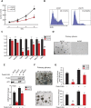
Zrf1 is required for neuroectodermal specification of ESCs. (A, left) Western blot for Zrf1 after infection of E14Tg2A ESCs with two different lentiviral shRNAs. (Right) Band intensity was quantified by using ImageJ software. Data represent the average of three independent experiments. (**) P < 0.01. (B) The relative mRNA expression of pluripotency markers after Zrf1 depletion. Values are normalized to Gapdh. Data represent the average of three independent experiments. (C) Mean fluorescent intensity of immunostained cells in shCtrl or shZrf1 teratomas quantified by using ImageJ software is depicted in the graph. Data represent the average of five selected teratomas (10 fields for each teratoma). (*) P < 0.05. (D) The relative mRNA expression of selected markers of three germ layers in entire teratomas. Values are normalized to Gapdh. The analysis was performed on five different teratomas. (*) P < 0.05. (E, left) FACS profile of Sox1-GFP expression ESCs grown in self-renewing condition (d0) and in N2B27 medium at day 5 (d5). (Right) The percentage of Sox1-GFP-positive cells at days 3 and 5 is reported in the graph. Data represent the average of three independent experiments. (**) P < 0.01. (F) Nestin immunostaining at day 5 in N2B27 medium. Bar, 200 μm. The percentage of positive (as compared with total DAPI [40,60-diamidino-2-phenylindole]-positive) cells is reported in the picture. Data represent three independent experiments (seven fields were counted for each experiment). (G) Western blot for β3-tubulin in shCtrl, shZrf1, and rescued cells (coexpressing shZrf1 and human Zrf1 cDNA tagged with 3xFlag) at day 5 in N2B27 medium.
Figure 2.
Figure 2.
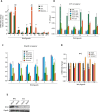
Zrf1 is required for generation of RGC-like NPCs from ESCs. (A) Scheme of generation of RGC-like NPCs. Briefly, ESCs were grown as cell aggregates (CAs) in medium supplemented with 10% serum. At day 4, RA was added to the medium to promote the generation of Pax6-positive RGC-like NPCs. At day 8, CAs were disaggregated, and RGC-like NPCs were harvested for the analysis. (B) Heat map illustrating the expression changes of selected NPC markers in shCtrl and shZrf1 CAs at day 6 of neural induction. Each lane corresponds to an independent biological sample. (C) The relative mRNA expression of NPC markers during generation of RGC-like NPCs. Values are normalized to Gapdh. Data represent the average of three independent experiments. (*) P < 0.05; (**) P < 0.01. (D) Zrf1 ChIP-qPCR (left panel) and Ring1B ChIP-qPCR (right panel) of selected NPC markers in ESCs (d0) and CAs at day 6 of neural specification (d6). Values are expressed as percentage of input. Data represent three different experiments. (**) P < 0.01. (E) Pax6 expression at mRNA (left panel) and protein (right panel) levels during NPC specification from ESCs. The graph represents data from three different experiments. (*) P < 0.05; (**) P < 0.01.
Figure 3.
Figure 3.
Zrf1 is required for self-renewal of ESC-derived NPCs. (A) Growth curve of shCtrl and shZrf1 NPCs and rescued NPCs. Data are representative of three independent experiments. (*) P < 0.05; (**) P < 0.01. (B) FACS plot of BrDU staining after incubation of 20 min in shCtrl and shZrf1 NPCs. Data are representative of three independent experiments. (C) The relative mRNA expression of several NPC markers in shCtrl and shZrf1 NPCs. Values are normalized to Gapdh. Data represent the average of three independent experiments. (*) P < 0.05; (**) P < 0.01. (D) Representative pictures of shCtrl and shZrf1 NPCs grown as spheres. Bars, 400 μm. (E, top panel) Western blot of Flag and Zrf1 expression in shCtrl and shZrf1 NPCs electroporated with Flag-tagged Pax6 cDNA. (Bottom panel) The relative mRNA expression levels of Pax6 in shCtrl and shZrf1 after Pax6 overexpression. (F) Representative pictures of shCtrl and shZrf1 NPCs after Pax6 overexpression grown as spheres. Bars, 250 μm. (G, top panel) Number of shCtrl and shZrf1 NPCs overexpressing Pax6 that gave rise to spheres over 1000 cells. (Bottom panel) Percentage of cells derived by disaggregation of primary spheres able to give rise to secondary spheres over the total number of cells. Data are representative of five different experiments. (**) P < 0.01.
Figure 4.
Figure 4.
A PRC1-dependent role of Zrf1 is involved in neurogenesis. (A) Overlap between Ring1B targets in ESCs and Zrf1 targets in NPCs. GO analysis reports enrichment in expression of total Ring1B and Zrf1 targets, common targets, and targets occupied by only Ring1B or Zrf1. GO analysis was performed by using Genomatix or DAVID software. (B) Overlap between Ring1B and Zrf1 targets in NPCs. GO analysis reports enrichment in expression of total Ring1B targets, common targets, and targets occupied by only Ring1B or Zrf1. GO analysis was performed by using Genomatix or DAVID software. (C) Ring1B ChIP-qPCR (top panel) and Zrf1 ChIP-qPCR (bottom panel) of selected genes in ESCs and NPCs. Values are reported as percentage of input. Data represent three different experiments. (**) P < 0.01.
Figure 5.
Figure 5.
Zrf1 controls the expression of several Wnt ligands in NPCs. (A) The relative mRNA expression of Zrf1-bound Wnt ligands in ESCs, NPCs, and post-mitotic neurons. Data are representative of three different experiments. (**) P < 0.01. (B) Zrf1 ChIP-qPCR of selected Wnt ligands in ESCs, NPCs, and post-mitotic neurons. Values are expressed as percentage of input. Data represent three different experiments. (**) P < 0.01. (C) Ring1B ChIP-qPCR of selected Wnt ligands in ESCs, NPCs, and post-mitotic neurons. Wnt4 was used as control. Values are expressed as percentage of input. Data represent three different experiments. (**) P < 0.01. (D) The relative mRNA expression level of Zrf1-bound Wnt ligands in NPCs as compared with ESCs. (**) P < 0.01. (E) Western blot of Wnt1 expression in shCtrl and shZrf1 ESCs and shCtrl and shZrf1 NPCs.
Figure 6.
Figure 6.
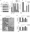
Zrf1-mediated control of Wnt signaling is essential for NPC self-renewal. (A, left) Western blot of active and total β-catenin, Pax6, and Zrf1 in shCtrl or shZrf1 NPCs treated or not with CHIR. (Right) Band intensity was quantified using the ImageJ software. Data represent the average of three independent experiments. (*) P < 0.05; (**) P < 0.01. (B) Pax6 mRNA expression levels in shCtrl and shZrf1 NPCs treated with CHIR or Wnt3a. (C) Representative pictures of spheres derived from shCtrl and shZrf1 NPCs treated with CHIR or Wnt3a. Bar, 150 μm. (D, top panel) Number of shCtrl and shZrf1 NPCs treated with CHIR, Wnt3a, or DMSO (as a control) that gave rise to primary spheres over 1000 cells. (Bottom panel) The percentage of cells derived by disaggregation of primary spheres able to give rise to secondary spheres over the total number of cells. Data are representative of five different experiments. (**) P < 0.01.
Figure 7.
Figure 7.
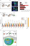
Zrf1 regulates the expression of Wnt ligands and self-renewal regulators in vivo. (A) Scheme of the experimental procedure. (B) Coronal hemisections of forebrains 48 h after electroporation with plasmids containing shCtrl or shZrf1. Bars, 100 μm. (C) Percentage of electroporated cells (GFP-positive) in five layers of the embryonic cortex 48 h after electroporation. (*) P < 0.05; (**) P < 0.01. (D) GFP-positive cells were isolated by FACS 48 h after electroporation with shCtrl, shZrf1, or shZrf1 + human Zrf1 cDNA in the VZ/subventricular zone (SVZ) of an E13.5 embryonic cortex. The relative mRNA expression of RGC markers and selected Wnt ligands is reported in the graph. Values are normalized to Gapdh. Data represent the average of three independent experiments (FACS-sorted cells from three different embryos were pooled together for each experiment). (*) P < 0.05; (**) P < 0.01. (E) Model: Zrf1 is essential to generate Pax6-positive NPCs from ESCs. In NPCs, Zrf1 directly regulates the expression of several Wnt ligand genes, which in turn initiate autocrine/paracrine canonical Wnt signaling, responsible for maintaining NPC self-renewal.

References

    1. Aboody K, Capela A, Niazi N, Stern JH, Temple S 2011. Translating stem cell studies to the clinic for CNS repair: Current state of the art and the need for a Rosetta stone. Neuron 70: 597–613 - PubMed
    1. Aloia L, Parisi S, Fusco L, Pastore L, Russo T 2010. Differentiation of embryonic stem cells 1 (Dies1) is a component of bone morphogenetic protein 4 (BMP4) signaling pathway required for proper differentiation of mouse embryonic stem cells. J Biol Chem 285: 7776–7783 - PMC - PubMed
    1. Aloia L, Di Stefano B, Di Croce L 2013. Polycomb complexes in stem cells and embryonic development. Development 140: 2525–2534 - PubMed
    1. Alvarez-Medina R, Le Dreau G, Ros M, Marti E 2009. Hedgehog activation is required upstream of Wnt signalling to control neural progenitor proliferation. Development 136: 3301–3309 - PubMed
    1. Aubert J, Dunstan H, Chambers I, Smith A 2002. Functional gene screening in embryonic stem cells implicates Wnt antagonism in neural differentiation. Nat Biotechnol 20: 1240–1245 - PubMed

Publication types

MeSH terms