. 2014 Jul;33(7):1101-11.
doi: 10.1007/s10096-013-2044-0. Epub 2014 Jan 22.

Bronchial microbiome of severe COPD patients colonised by Pseudomonas aeruginosa

Affiliations

Bronchial microbiome of severe COPD patients colonised by Pseudomonas aeruginosa

L Millares et al. Eur J Clin Microbiol Infect Dis. 2014 Jul.

Abstract

The bronchial microbiome in severe COPD during stability and exacerbation in patients chronically colonised by Pseudomonas aeruginosa (PA), has not been defined. Our objective was to determine the characteristics of the bronchial microbiome of severe COPD patients colonised and not colonised by P. aeruginosa and its changes during exacerbation. COPD patients with severe disease and frequent exacerbations were categorised according to chronic colonisation by P. aeruginosa. Sputum samples were obtained in stability and exacerbation, cultured, and analysed by 16S rRNA gene amplification and pyrosequencing. Sixteen patients were included, 5 of them showing chronic colonisation by P. aeruginosa. Pseudomonas genus had significantly higher relative abundance in stable colonised patients (p = 0.019), but no significant differences in biodiversity parameters were found between the two groups (Shannon, 3 (2-4) vs 3 (2-3), p = 0.699; Chao1, 124 (77-159) vs 140 (115-163), p = 0.364). In PA-colonised patients bronchial microbiome changed to a microbiome similar to non-PA-colonised patients during exacerbations. An increase in the relative abundance over 20 % during exacerbation was found for Streptococcus, Pseudomonas, Moraxella, Haemophilus, Neisseria, Achromobacter and Corynebacterium genera, which include recognised potentially pathogenic microorganisms, in 13 patients colonised and not colonised by P. aeruginosa with paired samples. These increases were not identified by culture in 5 out of 13 participants (38.5 %). Stable COPD patients with severe disease and PA-colonised showed a similar biodiversity to non-PA-colonised patients, with a higher relative abundance of Pseudomonas genus in bronchial secretions. Exacerbation in severe COPD patients showed the same microbial pattern, independently of previous colonisation by P. aeruginosa.


Figures
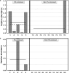
Fig. 1
Fig. 1
Relative abundance of Pseudomonas genus a under stability and b during exacerbation in PA-colonised and non-PA-colonised patients. Solid line represents the mean and dashed line the median
Fig. 2
Fig. 2
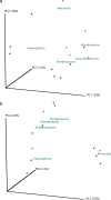
Principal coordinate analysis with Bray–Curtis dissimilarity index. a Samples from stability and b samples of exacerbation. Red dots represent colonised patients, and blue dots non-PA-colonised patients
Fig. 3
Fig. 3
Cluster dendrogram with Bray–Curtis dissimilarity index. Samples were combined depending on their clinical situation and clustered with the Bray–Curtis index, which takes values between 0 and 1 (0 meaning that samples share all the genera and 1 meaning samples do not share any). PS colonised by P. aeruginosa under stability; PE colonised by P. aeruginosa during exacerbation; S non-colonised by P. aeruginosa under stability; E non-colonised by P. aeruginosa during exacerbation)
Fig. 4
Fig. 4
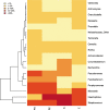
Heat map showing the most abundant genera in the four groups of samples. Columns represent the groups and rows the genera whose relative abundance is >1 % in at least one sample. The relative abundance of each genus is represented by the colour key
Fig. 5
Fig. 5
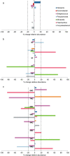
Percentage of change in the relative abundance of genera. Only genera with a percentage of change over 20 % in at least one patient are represented. a Percentage of variability in the genera observed in consecutive samples recovered in stable situation (n = 2); b Percentage of variability in exacerbation in patients colonised by P. aeruginosa using the baseline stability sample as the reference (n = 5); c Percentage of variability in exacerbation in non-PA-colonised patients (n = 7). Dark blue: Neisseria; orange: Achromobacter; purple: Streptococcus; light blue: Pseudomonas; green: Moraxella; pink: Haemophilus; grey: Corynebacterium

Similar articles

Cited by

References

    1. Hill AT, Campbell EJ, Hill SL, Bayley DL, Stockley RA. Association between airway bacterial load and markers of airway inflammation in patients with stable chronic bronchitis. Am J Med. 2000;109(4):288–295. doi: 10.1016/S0002-9343(00)00507-6. - DOI - PubMed
    1. Sethi S, Evans N, Grant BJ, Murphy TF. New strains of bacteria and exacerbations of chronic obstructive pulmonary disease. N Engl J Med. 2002;347(7):465–471. doi: 10.1056/NEJMoa012561. - DOI - PubMed
    1. Soler N, Torres A, Ewig S, Gonzalez J, Celis R, El-Ebiary M, Hernandez C, Rodriguez-Roisin R. Bronchial microbial patterns in severe exacerbations of chronic obstructive pulmonary disease (COPD) requiring mechanical ventilation. Am J Respir Crit Care Med. 1998;157(5 Pt 1):1498–1505. doi: 10.1164/ajrccm.157.5.9711044. - DOI - PubMed
    1. Eller J, Ede A, Schaberg T, Niederman MS, Mauch H, Lode H. Infective exacerbations of chronic bronchitis: relation between bacteriologic etiology and lung function. Chest. 1998;113(6):1542–1548. doi: 10.1378/chest.113.6.1542. - DOI - PubMed
    1. Rosell A, Monso E, Soler N, Torres F, Angrill J, Riise G, Zalacain R, Morera J, Torres A. Microbiologic determinants of exacerbation in chronic obstructive pulmonary disease. Arch Intern Med. 2005;165(8):891–897. doi: 10.1001/archinte.165.8.891. - DOI - PubMed

Publication types

Associated data

LinkOut - more resources