Detection of enthesitis in children with enthesitis-related arthritis: dolorimetry compared to ultrasonography
- PMID: 24449586
- PMCID: PMC3964147
- DOI: 10.1002/art.38197
Detection of enthesitis in children with enthesitis-related arthritis: dolorimetry compared to ultrasonography
Abstract
Objective: To evaluate the distribution of enthesitis and the accuracy of physical examination with a dolorimeter for the detection of enthesitis in children, using ultrasound (US) assessment as the reference standard.
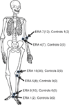
Methods: We performed a prospective cross-sectional study of 30 patients with enthesitis-related arthritis (ERA) and 30 control subjects. The following tendon insertion sites were assessed by standardized physical examination with a dolorimeter and US: common extensor on the lateral humeral epicondyle, common flexor on the medial humeral epicondyle, quadriceps at the superior patella, patellar ligament at the inferior patella, Achilles, and plantar fascia at the calcaneus.
Results: Abnormal findings on US were detected most commonly at the insertion of the quadriceps (30% [18 of 60 sites]), common extensor (12% [7 of 60]), and Achilles (10% [6 of 60]) tendons. The intrarater reliability of US (kappa statistic) was 0.78 (95% confidence interval [95% CI] 0.63-0.93), and the interrater reliability was 0.81 (95% CI 0.67-0.95). Tenderness as detected by standardized dolorimeter examination had poor positive predictive value for US-confirmed enthesitis. In comparison to controls, patients with ERA reported more pain and had lower pain thresholds at every site, including control sites (P < 0.001 for all comparisons). The interrater reliability of dolorimeter examination for detection of enthesitis was low (κ = 0.49 [95% CI 0.33-0.65]).
Conclusion: Compared to US, standardized dolorimeter examination for the detection of enthesitis in children has poor accuracy and reliability. The decreased pain threshold of ERA patients likely contributed to the limited accuracy of the physical examination findings. Further research regarding the utility of US for identifying enthesitis at diagnosis of juvenile idiopathic arthritis, accurately predicting disease progression, and guiding therapeutic decisions is warranted.
Copyright © 2014 by the American College of Rheumatology.
Figures


Similar articles
-
Enthesitis in an inception cohort of enthesitis-related arthritis.Arthritis Care Res (Hoboken). 2011 Sep;63(9):1307-12. doi: 10.1002/acr.20508. Arthritis Care Res (Hoboken). 2011. PMID: 21618453
-
Ultrasound of the joints and entheses in healthy children.Pediatr Radiol. 2015 Aug;45(9):1344-54. doi: 10.1007/s00247-015-3313-0. Epub 2015 Mar 6. Pediatr Radiol. 2015. PMID: 25744571
-
[Ultrasonic Features of Enthesitis: a Comparison Between Patients with Psoriasis Vulgaris and Patients with Psoriatic Arthritis].Sichuan Da Xue Xue Bao Yi Xue Ban. 2017 Jul;48(4):589-594. Sichuan Da Xue Xue Bao Yi Xue Ban. 2017. PMID: 28752980 Chinese.
-
Validity of ultrasonography in detecting enthesitis in children: A systematic literature review.Joint Bone Spine. 2023 Jul;90(4):105538. doi: 10.1016/j.jbspin.2023.105538. Epub 2023 Feb 6. Joint Bone Spine. 2023. PMID: 36754113
-
Enthesitis-related arthritis.Clin Rheumatol. 2015 Nov;34(11):1839-46. doi: 10.1007/s10067-015-3029-4. Epub 2015 Aug 2. Clin Rheumatol. 2015. PMID: 26233720 Review.
Cited by
-
Detection of inflammation by whole-body MRI in young people with juvenile idiopathic arthritis.Rheumatology (Oxford). 2024 Sep 1;63(SI2):SI207-SI214. doi: 10.1093/rheumatology/keae039. Rheumatology (Oxford). 2024. PMID: 38244609 Free PMC article.
-
Ultrasound in juvenile idiopathic arthritis.Pediatr Rheumatol Online J. 2016 May 27;14(1):33. doi: 10.1186/s12969-016-0096-2. Pediatr Rheumatol Online J. 2016. PMID: 27234966 Free PMC article. Review.
-
Outcomes in Juvenile-Onset Spondyloarthritis.Front Med (Lausanne). 2021 May 28;8:680916. doi: 10.3389/fmed.2021.680916. eCollection 2021. Front Med (Lausanne). 2021. PMID: 34124112 Free PMC article. Review.
-
Contribution of Ultrasound in Current Practice for Managing Juvenile Idiopathic Arthritis.J Clin Med. 2022 Dec 22;12(1):91. doi: 10.3390/jcm12010091. J Clin Med. 2022. PMID: 36614888 Free PMC article. Review.
-
Advances in the Diagnosis and Treatment of Enthesitis-Related Arthritis.Children (Basel). 2023 Oct 2;10(10):1647. doi: 10.3390/children10101647. Children (Basel). 2023. PMID: 37892310 Free PMC article. Review.
References
-
- Sherry DD, Sapp LR. Enthesalgia in childhood: site-specific tenderness in healthy subjects and in patients with seronegative enthesopathic arthropathy. J Rheumatol. 2003;30(6):1335–1340. - PubMed
-
- D'Agostino MA, Said-Nahal R, Hacquard-Bouder C, Brasseur JL, Dougados M, Breban M. Assessment of peripheral enthesitis in the spondylarthropathies by ultrasonography combined with power Doppler: a cross-sectional study. Arthritis Rheum. 2003;48(2):523–533. - PubMed
-
- Sprott H, Jeschonneck M, Grohmann G, Hein G. [Microcirculatory changes over the tender points in fibromyalgia patients after acupuncture therapy (measured with laser-Doppler flowmetry)] Wien Klin Wochenschr. 2000;112(13):580–586. - PubMed
-
- D'Agostino MA, Breban M, Brasseur JL, Canale M, Magaro M. Ultrasonographic (US) assessment of spondyloarthropathy (SpA) enthesitis, displays vascularization change specific of inflammatory process [abstract] Arthritis Rheum. 2001;44(Suppl 9):S95.
-
- Aydin SZ, Karadag O, Filippucci E, Atagunduz P, Akdogan A, Kalyoncu U, et al. Monitoring Achilles enthesitis in ankylosing spondylitis during TNF-alpha antagonist therapy: an ultrasound study. Rheumatology (Oxford) 49(3):578–582. - PubMed
Publication types
MeSH terms
Grants and funding
LinkOut - more resources
Full Text Sources
Other Literature Sources
Medical

