. 2014 Jan 21;5(1):e00775-13.
doi: 10.1128/mBio.00775-13.

Structural basis for the inhibition of the chromatin repressor BAHD1 by the bacterial nucleomodulin LntA

Structural basis for the inhibition of the chromatin repressor BAHD1 by the bacterial nucleomodulin LntA

Alice Lebreton et al. mBio. .

Abstract

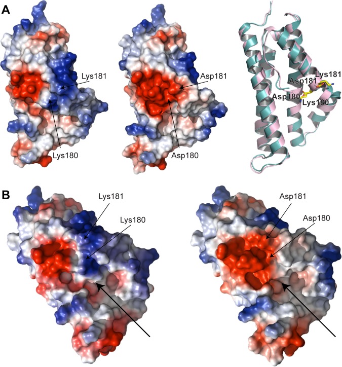
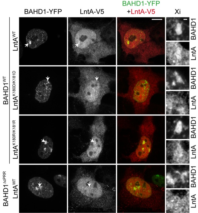
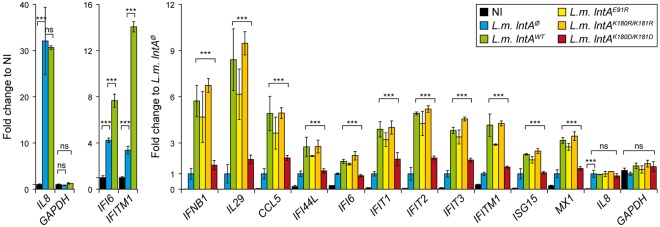
The nucleus has emerged as a key target for nucleomodulins, a family of effectors produced by bacterial pathogens to control host transcription or other nuclear processes. The virulence factor LntA from Listeria monocytogenes stimulates interferon responses during infection by inhibiting BAHD1, a nuclear protein involved in gene silencing by promoting heterochromatin formation. So far, whether the interaction between LntA and BAHD1 is direct and sufficient for inhibiting BAHD1 activity is unknown. Here, we functionally characterized the molecular interface between the two proteins in vitro and in transfected or infected human cells. Based on the known tridimensional structure of LntA, we identified a dilysine motif (K180/K181) in the elbow region of LntA and a central proline-rich region in BAHD1 as crucial for the direct LntA-BAHD1 interaction. To better understand the role played by the dilysine motif in the functionality of LntA, we solved the crystal structure of a K180D/K181D mutant to a 2.2-Å resolution. This mutant highlights a drastic redistribution of surface charges in the vicinity of a groove, which likely plays a role in nucleomodulin target recognition. Mutation of the strategic dilysine motif also abolished the recruitment of LntA to BAHD1-associated nuclear foci and impaired the LntA-mediated stimulation of interferon responses upon infection. Last, the strict conservation of residues K180 and K181 in LntA sequences from 188 L. monocytogenes strains of different serotypes and origins further supports their functional importance. Together, these results provide structural and functional details about the mechanism of inhibition of an epigenetic factor by a bacterial nucleomodulin.

Importance: Pathogens have evolved various strategies to deregulate the expression of host defense genes during infection, such as targeting nuclear proteins. LntA, a secreted virulence factor from the bacterium Listeria monocytogenes, stimulates innate immune responses by inhibiting a chromatin-associated repressor, BAHD1. This study reveals the structural features of LntA required for BAHD1 inhibition. LntA interacts directly with a central domain of BAHD1 via a surface patch of conserved positive charges, located nearby a groove on the elbow region of LntA. By demonstrating that this patch is required for LntA function, we provide a better understanding of the molecular mechanism allowing a bacterial pathogen to control host chromatin compaction and gene expression.


Figures

FIG 1
FIG 1
LntA interacts in vitro with BAHD1187–428. (A) Primary structure of BAHD1. GST-BAHD1187–428 and GST-BAHD1239–361 fusions were tested for interaction with LntA. cPRR, central proline-rich region; NLS, nuclear localization signal; BAH, bromo-adjacent homology domain. (B) Gel filtration experiment. Purified LntA, GST-BAHD1187–428, or GST-BAHD1239–361, incubated alone or coincubated for 3 h at 4°C, were injected on a Superdex 200 column. Elution fractions (lanes 1 to 9) were analyzed by SDS-PAGE followed by Coomassie blue staining. Elution fractions of single proteins are shown in the first three lines; those of coincubated proteins are highlighted in the gels labeled a and b. *, coelution of LntA and GST-BAHD1187–428. (C) 6His-LntA was coexpressed in E. coli with GST-BAHD1187–428 or GST-BAHD1239–361. Input and eluted fractions from pulldown assays on His-Trap or GST-Trap columns were analyzed by immunoblotting against the GST or the 6His tag.
FIG 2
FIG 2
The surface of LntA displays two major basic patches and one acidic patch. (A) LntA folds into a compact helical structure that is stabilized by a ladder of hydrophobic residues, indicated as yellow sticks. The thermal-motion color scale (B-factor values) indicates that the body formed by H1, H2, and H3 is the less mobile region and that the H4-H5 elbow displays more flexibility. Nter, N terminus. (B) The three main charged patches targeted by mutagenesis are shown (green circles) on the electrostatic surface views of LntA. Basic regions are in blue and acidic regions in red. Patch-forming residues are shown as sticks.
FIG 3
FIG 3
Positive charges in patch 3 of LntA are critical for interaction with BAHD1. (A) 6His-tagged LntAWT and LntA mutants coexpressed with GST-BAHD1187–428 in E. coli were used for GST pulldown assays. 6His-LntAWT alone and 6His-LntAWT coexpressed with GST-BAHD1239–361 were used as negative controls. Proteins were detected by immunoblotting against the tags. (B) GST-tagged LntAWT, LntA mutants, or InlC bound to glutathione-Sepharose was incubated with chromatin extracts containing full-length BAHD1-V5 or CFP-V5 as a negative control. V5-tagged fusions were detected by immunoblotting. GST-fused baits were detected by colloidal Coomassie blue staining.
FIG 4
FIG 4
Structural comparison of LntAWT and LntAK180D/K181D. (A) Comparison of charged patches on the surfaces of LntAWT (left) and LntAK180D/K181D (middle). On the right, a ribbon overlay shows LntAWT in light teal and LntAK180D/K181D in light pink. The mutated residues are indicated as sticks. (B) Arrows indicate the direction of an elongated groove in the structures, which ends with the basic patch affected by the mutations.
FIG 5
FIG 5
The K180/K181 dilysine motif in LntA and the proline-rich region in BAHD1 are required for the colocalization of the two proteins in human cells. 293FT cells were cotransfected for 48 h with WT or mutant versions of pcDNA3-lntA-V5 and pEYFP-N1-BAHD1. For lntA-V5, alleles resulted in WT, K180D/K181D, or K180R/K181R proteins. For BAHD1-YFP, alleles resulted in WT or ∆cPRR (i.e., missing residues 239 to 361) proteins. LntA-V5 and BAHD1-YFP were detected by immunofluorescence confocal microscopy. Arrows indicate the position of an Xi. A magnification of these regions with enhanced contrasts is shown on the left. Bars, 10 µm for the cell view and 1 µm for magnified Xi regions.
FIG 6
FIG 6
Stimulation of ISG expression by LntA during infection is impaired by mutation of the K180/K181 dilysine motif. Innate immune gene expression was quantified by RT-qPCR in noninfected LoVo cells (NI) or cells infected for 18 h with L. monocytogenes lntA, lntAWT, lntAK180R/K181R, or lntAK180D/K181D (left). Induction of the expression of interleukin 8 (IL-8), IFI6, and IFITM1 by L. monocytogenes lntA and L. monocytogenes lntAWT, compared to uninfected cells (right). LntA-dependent induction of ISGs, monitored by normalization to cells infected with L. monocytogenes lntA. Error bars indicate standard deviations. The reference gene for normalization was YWHAZ; GAPDH was a control invariant gene. Statistical analysis was performed using analysis of variance (ANOVA) followed by the Tukey-Kramer posttest. ns, nonsignificant; ***, P < 0.001.

Similar articles

Cited by

References

    1. Allerberger F, Wagner M. 2010. Listeriosis: a resurgent foodborne infection. Clin. Microbiol. Infect. 16:16–23. 10.1111/j.1469-0691.2009.03109.x - DOI - PubMed
    1. Stavru F, Archambaud C, Cossart P. 2011. Cell biology and immunology of Listeria monocytogenes infections: novel insights. Immunol. Rev. 240:160–184. 10.1111/j.1600-065X.2010.00993.x - DOI - PubMed
    1. Zenewicz LA, Shen H. 2007. Innate and adaptive immune responses to Listeria monocytogenes: a short overview. Microbes Infect. 9:1208–1215. 10.1016/j.micinf.2007.05.008 - DOI - PMC - PubMed
    1. Camejo A, Carvalho F, Reis O, Leitão E, Sousa S, Cabanes D. 2011. The arsenal of virulence factors deployed by Listeria monocytogenes to promote its cell infection cycle. Virulence 2:379–394. 10.4161/viru.2.5.17703 - DOI - PubMed
    1. Cossart P. 2011. Illuminating the landscape of host-pathogen interactions with the bacterium Listeria monocytogenes. Proc. Natl. Acad. Sci. U. S. A. 108:19484–19491. 10.1073/pnas.1112371108 - DOI - PMC - PubMed

Publication types

Associated data

LinkOut - more resources