Randomized Controlled Trial
. 2014 Apr 11;114(8):1292-301.
doi: 10.1161/CIRCRESAHA.114.302854. Epub 2014 Jan 21.

Does transendocardial injection of mesenchymal stem cells improve myocardial function locally or globally?: An analysis from the Percutaneous Stem Cell Injection Delivery Effects on Neomyogenesis (POSEIDON) randomized trial

Affiliations
Randomized Controlled Trial

Does transendocardial injection of mesenchymal stem cells improve myocardial function locally or globally?: An analysis from the Percutaneous Stem Cell Injection Delivery Effects on Neomyogenesis (POSEIDON) randomized trial

Viky Y Suncion et al. Circ Res. .

Abstract

Rationale: Transendocardial stem cell injection (TESI) with mesenchymal stem cells improves remodeling in chronic ischemic cardiomyopathy, but the effect of the injection site remains unknown.

Objective: To address whether TESI exerts its effects at the site of injection only or also in remote areas, we hypothesized that segmental myocardial scar and segmental ejection fraction improve to a greater extent in injected than in noninjected segments.

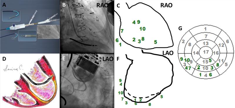
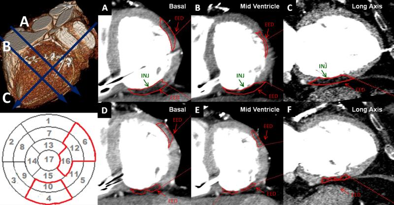
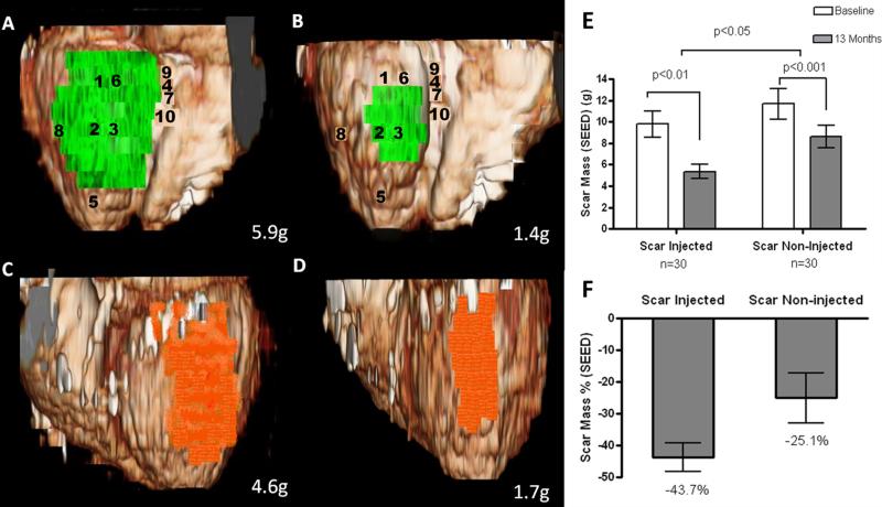
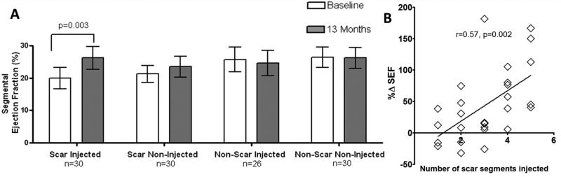
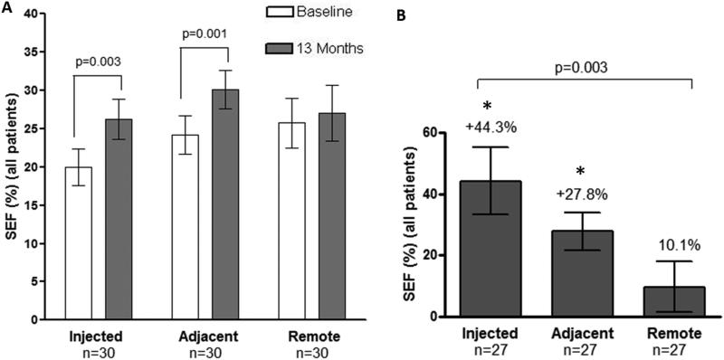
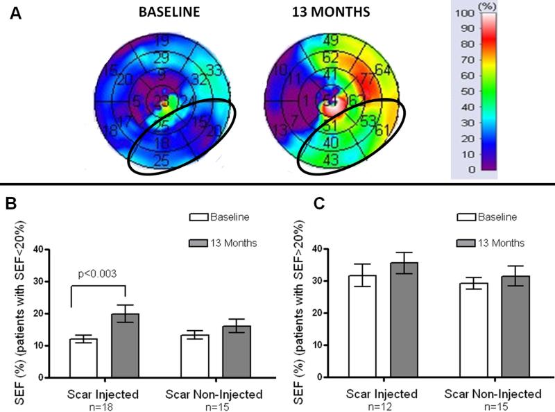
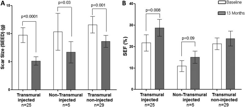
Methods and results: Biplane ventriculographic and endocardial tracings were recorded. TESI was guided to 10 sites in infarct-border zones. Sites were mapped according to the 17-myocardial segment model. As a result, 510 segments were analyzed in 30 patients before and 13 months after TESI. Segmental early enhancement defect (a measure of scar size) was reduced by TESI in both injected (-43.7 ± 4.4%; n=95; P<0.01) and noninjected segments (-25.1 ± 7.8%; n=148; P<0.001; between-group comparison P<0.05). Conversely, segmental ejection fraction (a measure of contractile performance) improved in injected scar segments (19.9 ± 3.3-26.3 ± 3.5%; P=0.003) but not in noninjected scar segments (21.3 ± 2.6-23.5 ± 3.2%; P=0.20; between-group comparison P<0.05). Furthermore, segmental ejection fraction in injected scar segments improved to a greater degree in patients with baseline segmental ejection fraction <20% (12.1 ± 1.2-19.9 ± 2.7%; n=18; P=0.003), versus <20% (31.7 ± 3.4-35.5 ± 3.3%; n=12; P=0.33, between-group comparison P<0.0001).

Conclusions: These findings illustrate a dichotomy in regional responses to TESI. Although scar size reduction was evident in all scar segments, scar size reduction and ventricular functional responses preferentially occurred at the sites of TESI versus non-TESI sites. Furthermore, improvement was greatest when segmental left ventricular dysfunction was severe.

Keywords: cells; magnetic resonance imaging; myocardial infarction; tomography.


Figures

Figure 1
Figure 1. Allocation of 10 injections using biplane ventriculography during Transendocardial Stem Cell Injection
(Panels A and D) Biocardia helical infusion catheter and schematic depiction of an intraendocardial injection, respectively. (Panels B and E) Biplane fluoroscopy was used for navigation. (Panels C and F) Every injection site was marked in two projections using ventriculographic diastolic contours and translated onto a 17-segment polar map (Panel G) to identify all segments that received intramyocardial injections of MSCs.
Figure 2
Figure 2. Greater reduction in scar injected segments
(Upper left Panel) 3D reconstruction at baseline and MDCT images with 20HU below the normal myocardium density at (Panel A) basal short-axis, (Panel B) mid-ventricle short-axis and (Panel C) long-axis representing EED areas delineated in red. (Lower left Panel) Polar map reconstruction with EED delineated in red. Scar mass (EED) at baseline (Panels A-C) has a greater reduction in the injected segments (marked by INJ) located in inferior segments at basal, mid-ventricle and long-axis, from 5.9g to 1.4g compared to (Panels D-F) non-injected lateral segments at basal, mid-ventricle and long-axis images, from 4.6g to 1.7g thirteen months after MSCs.
Figure 3
Figure 3. Volume rendered 3D reformats of left ventricle with color encoding of scar tissue
(Panels A and B) Scar mass (green) of inferior segments treated by TESI at baseline and 13-months post TESI, respectively (numbers represent sites of injection). (Panels C and D) Scar mass (orange) of lateral segments not treated by TESI at baseline and at 13-month follow up, respectively. Actual scar mass (grams) is depicted in the lower right corner of each panel. 3D-recontructions in this figure correspond to SEED measurements of the same patient as in Figure 2 and Online Video I. (Panels E and F) Absolute values and %changes of scar mass obtained by segmental imaging analysis approach. When considering the autologous and allogeneic groups combined, there is greater scar size reduction in the “scar-injected” segments (−43.7±4.4%, from 9.8±1.2 to 5.4±0.7g, n=30, p<0.01), compared to the “scar-non-injected” segments (−25.1±7.8%, from 11.7±1.4 to 8.6±1.0g, n=30, p<0.001; between group comparison “scar-injected” vs. “scar-non-injected” p<0.05).
Figure 4
Figure 4. Restoration in contractility (SEF) and association between number of injected segments vs. SEF
(Panel A) Greater improvement in SEF was observed in patients with more scar (SEED) injected segments, when considering the autologous and allogeneic groups combined, “scar-injected” segments SEF improved from 19.9±3.3% to 26.3±3.5% at 13-months after TESI (p=0.003). Conversely, SEF did not significantly improve when “scar-non-injected” segments were evaluated. The “non-scar-injected” and “non-scar-non-injected”segments did not demonstrate changes in SEF at 13-months. (Panel B) Pearson correlation coefficient r=0.57, 95% CI, 0.23 to 0.78; two-tailed p value=0.002.
Figure 5
Figure 5. Radius of Activity for Transendocardial Stem Cell Injections (TESI) with Mesenchymal Stem Cells in Segmental Ejection Fraction (SEF%)
(Panel A) Improvement in contractility was observed in “scar-injected” (from 19.9±3.3% at screening to 26.3±3.5% at 13-month after TESI (p=0.003), and “adjacent” segments (from 24.2±2.6% to 30.1±2.6%, n=30, p=0.001). Conversely, the “remote” segments did not have changes in contractility (15.8 ±12.4% from 25.8±3.3% to 27.1±3.7%, n=30, p=0.4); between group comparison p>0.05). (Panel B) Percent changes from baseline in SEF showed changes by +44.3%±11.2% in “scar-injected”, by +27.8±6.4% in “adjacent” and by 10.1±8.9% in the “remote” segments (one way analysis of variance p=0.003. Dunn's posttest *p<0.05 in “scar-injected” vs. “remote” and “adjacent” vs. “remote”).
Figure 6
Figure 6. Improvement in contractility is particularly evident in the highly dysfunctional segments
(Panel A) SEF<20%, scar-injected segments (baseline) which are encircled with an ellipse in the 17-segment polar map with their corresponding improvement after injection of mesenchymal stem cells. (Panel B) Represents the changes in SEF with highly dysfunctional segments (SEF<20%), the “scar- injected” SEF was 12.1±1.23% at baseline and improved to 19.9±2.69% (p=0.003) at 13-months after TESI (n=18 patients). In this subgroup analysis, the “scar-non-injected” SEF (n=15 patients) showed a trend of increase from 13.3±1.3% to 16.1±2.13%, (p=0.05).(Panel C) Shows changes in segments with SEF>20%.
Figure 7
Figure 7. Dual effects of MSCs, global scar size reduction but restoration of contractility only in scar-injected segments, regardless of transmurality
(Panel A) Similar magnitude of change in SEED of the “scar-injected” and “non-injected” with a transmural extent of >50% (from 9.8±1.3 to 5.2±0.7g, n=25, p<0.0001), and <50% (from 10.4±3.3 to 6.7±5.8g, n= 5, p=0.03). (Panel B) Similar magnitude of change in SEF only in the “scar-injected” with a transmural extent of >50% (from 21.8±3.8% to 28.8±4.0%, n=25, p=0.008) and <50% (from 11.0±2.6 to 15.1±2.9, n=5, p=0.09).

Comment in

References

    1. Schuleri KH, Feigenbaum GS, Centola M, Weiss ES, Zimmet JM, Turney J, Kellner J, Zviman MM, Hatzistergos KE, Detrick B, Conte JV, McNiece I, Steenbergen C, Lardo AC, Hare JM. Autologous mesenchymal stem cells produce reverse remodelling in chronic ischaemic cardiomyopathy. Eur Heart J. 2009;30:2722–2732. - PMC - PubMed
    1. Amado LC, Saliaris AP, Schuleri KH, St John M, Xie JS, Cattaneo S, Durand DJ, Fitton T, Kuang JQ, Stewart G, Lehrke S, Baumgartner WW, Martin BJ, Heldman AW, Hare JM. Cardiac repair with intramyocardial injection of allogeneic mesenchymal stem cells after myocardial infarction. Proc Natl Acad Sci U S A. 2005;102:11474–11479. - PMC - PubMed
    1. Williams AR, Trachtenberg B, Velazquez DL, McNiece I, Altman P, Rouy D, Mendizabal AM, Pattany PM, Lopera GA, Fishman J, Zambrano JP, Heldman AW, Hare JM. Intramyocardial stem cell injection in patients with ischemic cardiomyopathy: Functional recovery and reverse remodeling. Circ Res. 2011;108:792–796. - PMC - PubMed
    1. Hare JM, Fishman JE, Gerstenblith G, DiFede Velazquez DL, Zambrano JP, Suncion VY, Tracy M, Ghersin E, Johnston PV, Brinker JA, Breton E, Davis-Sproul J, Schulman IH, Byrnes J, Mendizabal AM, Lowery MH, Rouy D, Altman P, Wong Po Foo C, Ruiz P, Amador A, Da Silva J, McNiece IK, Heldman AW, George R, Lardo A. Comparison of allogeneic vs autologous bone marrow-derived mesenchymal stem cells delivered by transendocardial injection in patients with ischemic cardiomyopathy: The poseidon randomized trial. JAMA : the journal of the American Medical Association. 2012;308:2369–2379. - PMC - PubMed
    1. Dib N, Menasche P, Bartunek JJ, Zeiher AM, Terzic A, Chronos NA, Henry TD, Peters NS, Fernandez-Aviles F, Yacoub M, Sanborn TA, Demaria A, Schatz RA, Taylor DA, Fuchs S, Itescu S, Miller LW, Dinsmore JH, Dangas GD, Popma JJ, Hall JL, Holmes DR., Jr. Recommendations for successful training on methods of delivery of biologics for cardiac regeneration: A report of the international society for cardiovascular translational research. JACC Cardiovasc Interv. 2010;3:265–275. - PubMed

Publication types

MeSH terms