. 2014 Feb 4;111(5):1807-12.
doi: 10.1073/pnas.1321852111. Epub 2014 Jan 21.

Nonautonomous contact guidance signaling during collective cell migration

Affiliations

Nonautonomous contact guidance signaling during collective cell migration

Camila Londono et al. Proc Natl Acad Sci U S A. .

Abstract

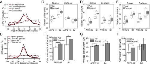
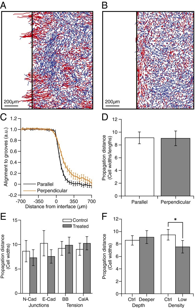
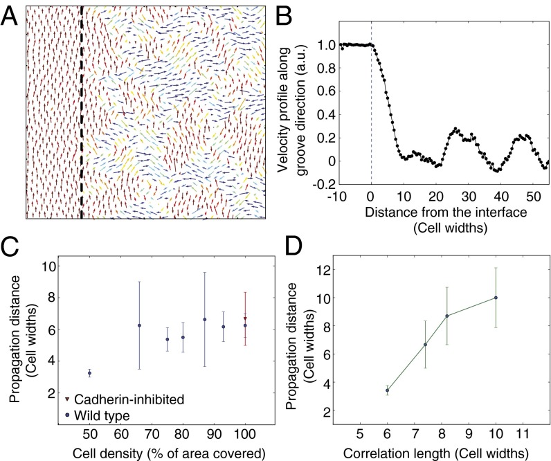
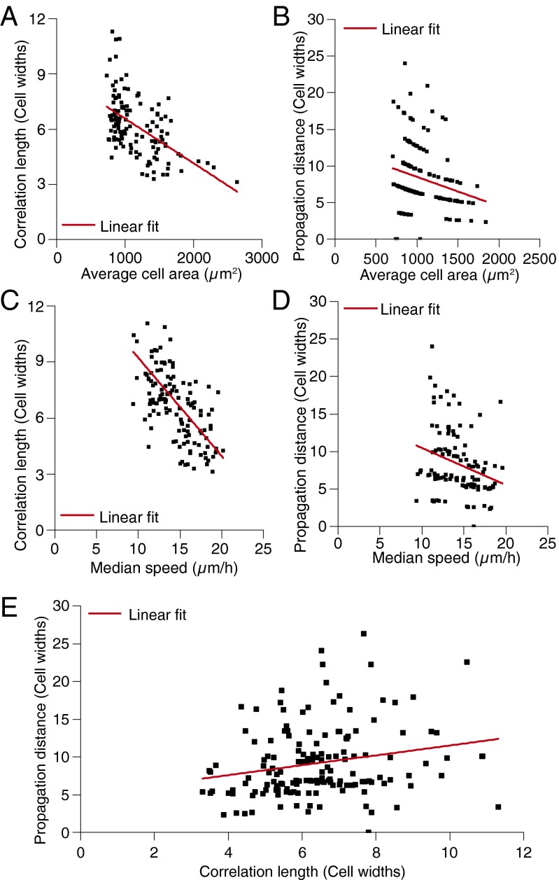
Directed migration of groups of cells is a critical aspect of tissue morphogenesis that ensures proper tissue organization and, consequently, function. Cells moving in groups, unlike single cells, must coordinate their migratory behavior to maintain tissue integrity. During directed migration, cells are guided by a combination of mechanical and chemical cues presented by neighboring cells and the surrounding extracellular matrix. One important class of signals that guide cell migration includes topographic cues. Although the contact guidance response of individual cells to topographic cues has been extensively characterized, little is known about the response of groups of cells to topographic cues, the impact of such cues on cell-cell coordination within groups, and the transmission of nonautonomous contact guidance information between neighboring cells. Here, we explore these phenomena by quantifying the migratory response of confluent monolayers of epithelial and fibroblast cells to contact guidance cues provided by grooved topography. We show that, in both sparse clusters and confluent sheets, individual cells are contact-guided by grooves and show more coordinated behavior on grooved versus flat substrates. Furthermore, we demonstrate both in vitro and in silico that the guidance signal provided by a groove can propagate between neighboring cells in a confluent monolayer, and that the distance over which signal propagation occurs is not significantly influenced by the strength of cell-cell junctions but is an emergent property, similar to cellular streaming, triggered by mechanical exclusion interactions within the collective system.

Keywords: correlation length; emergent behavior; group coordination; mechanical signal propagation.


Conflict of interest statement

The authors declare no conflict of interest.

Figures

Fig. 1.
Fig. 1.
Quantification of cell migration and coordination behavior on flat and grooved substrates. Cell migration characteristics of ARPE-19 and BJ cells at sparse and confluent cell densities on flat versus grooved substrates. (A and B) Distribution of cell migration direction on flat versus grooved substrates for (A) ARPE-19 and (B) BJ cells. The peak created by the grooved substrates between −25° and 25° indicates cell migration guidance. (C) Cell speed was significantly increased by grooves in sparse ARPE-19 and BJ cells, but not in confluent cell sheets of ARPE-19 or BJ cells. (D) Cell velocity was significantly increased by grooves in sparse ARPE-19 and BJ cells and in confluent cell sheets of ARPE-19 and BJ. (E) Cell persistence was significantly increased by grooves in sparse ARPE-19 and in confluent cell sheets of ARPE-19 and BJ but not significantly different in sparse BJ cells. (F) Grooves significantly increased the percentage of cells participating in streams of width greater than 40 µm for both ARPE-19 and BJ cells. (G) Grooves significantly increased stream width in both ARPE-19 and BJ cells. (H) Grooves significantly increased correlation length in ARPE-19 but not BJ cells. Error bars represent 95% confidence intervals.
Fig. 2.
Fig. 2.
Quantification of guidance propagation from a topographic feature. Guidance propagates about nine cells. Groove depth, adherens junctions, and myosin activity do not affect propagation distance. (A and B) Migration tracks of cells located on the flat region at start of the experiment on surfaces with (A) grooves perpendicular or (B) grooves parallel to the interface. Tracks are colored according to their angles [guided (red) versus nonguided (blue)]. (C) Quantification of alignment of cell migration direction with groove direction as a function of distance from the interface for grooves parallel or perpendicular to the interface. (D) Propagation distance from the interface, quantified as the distance at which the curves in C reach 0.321 (i.e., cells no longer move within 25° of the groove direction) for grooves parallel and perpendicular to the interface. (E) Propagation of guidance signals from the interface for parallel grooves in GFP-N-cadherin overexpressing ARPE-19 cells, anti–E-cadherin-treated ARPE-19 cells, blebbistain (BB)-treated cells, and calyculin A (CalA)-treated cells. No significant difference in signal propagation distance was observed between treated and control cells. Measurements were made on at least 19 wells from at least three independent experiments. (F) Propagation distance from the interface measured for parallel grooves of different depths and for cell sheets at different densities. No significant difference in signal propagation distance was observed between deep and shallow grooves. Sheets with a high cell density did, however, show a significantly higher propagation distance than cells in low-density sheets.
Fig. 3.
Fig. 3.
Propagation distance predictions from computational modeling. (A) Cell velocity field from computational model. Cells to the left of the interface (indicated by the dashed line) were biased to move vertically, mimicking cells on grooves. No bias was applied to cells to the right of the interface. Arrows are placed at the center of each cell within the sheet. Colors indicate the direction of migration. (B) Mean vertical velocity as a function of the distance from the interface for the parallel grooves case. The data are normalized so that the mean vertical velocity on the grooves is 1. The propagation distance is defined by the intersection between the curve and 0.321. (C) Propagation as a function of cell density for wild-type (blue) and cadherin-inhibited (red) cells. (D) Propagation distance as a function of the correlation of the cell population. According to the model, a correlation exists between propagation distance and correlation length. Error bars represent 95% confidence intervals.
Fig. 4.
Fig. 4.
Identifying relationships between propagation distance, correlation length, cell density, and cell speed from in vitro data. (A) Correlation length as a function of average cell area measured from cells on flat substrate regions showing a significant correlation (R = 0.538 versus Rsignificant = 0.195 for n = 127). (B) Signal propagation distance as a function of average cell area measured at the interface showing a significant correlation (R = 0.241 versus Rsignificant = 0.138 for n = 154). Propagation distance being measured in 40-µm bins results in the observed banding/segregation of the data. (C) Correlation length as a function of median cell speed measured from cells on flat substrate regions showing a significant correlation (R = 0.623 versus Rsignificant = 0.195 for n = 127). (D) Signal propagation distance as a function of median cell speed (speed measurements made on flat substrate regions) showing a significant correlation (R = 0.284 versus Rsignificant = 0.195 for n = 116). (E) Propagation distance as a function of correlation length (correlation length measured from cells on flat substrate regions) showing a significant correlation (R = 0.214 versus Rsignificant = 0.138 for n = 153).

References

    1. McMahon A, Reeves GT, Supatto W, Stathopoulos A. Mesoderm migration in Drosophila is a multi-step process requiring FGF signaling and integrin activity. Development. 2010;137(13):2167–2175. - PMC - PubMed
    1. Petrie RJ, Doyle AD, Yamada KM. Random versus directionally persistent cell migration. Nat Rev Mol Cell Biol. 2009;10(8):538–549. - PMC - PubMed
    1. Calve S, Simon H-G. Biochemical and mechanical environment cooperatively regulate skeletal muscle regeneration. FASEB J. 2012;26(6):2538–2545. - PMC - PubMed
    1. Friedl P, Gilmour D. Collective cell migration in morphogenesis, regeneration and cancer. Nat Rev Mol Cell Biol. 2009;10(7):445–457. - PubMed
    1. Trepat X, Fredberg JJ. Plithotaxis and emergent dynamics in collective cellular migration. Trends Cell Biol. 2011;21(11):638–646. - PMC - PubMed

Publication types