. 2014 Feb 4;111(5):1825-30.
doi: 10.1073/pnas.1319402111. Epub 2014 Jan 21.

Functional map of arrestin-1 at single amino acid resolution

Affiliations

Functional map of arrestin-1 at single amino acid resolution

Martin K Ostermaier et al. Proc Natl Acad Sci U S A. .

Abstract

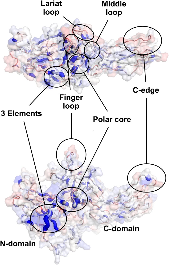
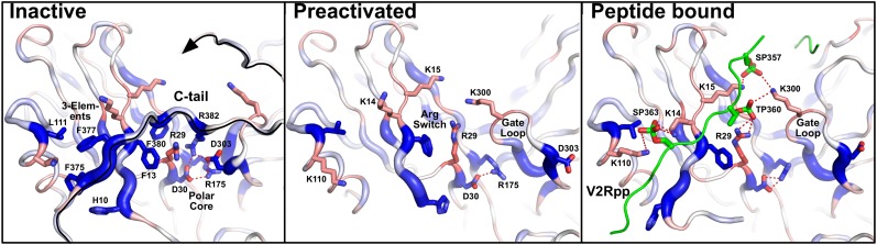
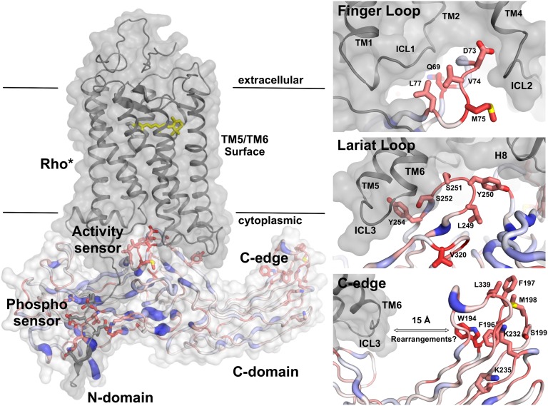
Arrestins function as adapter proteins that mediate G protein-coupled receptor (GPCR) desensitization, internalization, and additional rounds of signaling. Here we have compared binding of the GPCR rhodopsin to 403 mutants of arrestin-1 covering its complete sequence. This comprehensive and unbiased mutagenesis approach provides a functional dimension to the crystal structures of inactive, preactivated p44 and phosphopeptide-bound arrestins and will guide our understanding of arrestin-GPCR complexes. The presented functional map quantitatively connects critical interactions in the polar core and along the C tail of arrestin. A series of amino acids (Phe375, Phe377, Phe380, and Arg382) anchor the C tail in a position that blocks binding of the receptor. Interaction of phosphates in the rhodopsin C terminus with Arg29 controls a C-tail exchange mechanism in which the C tail of arrestin is released and exposes several charged amino acids (Lys14, Lys15, Arg18, Lys20, Lys110, and Lys300) for binding of the phosphorylated receptor C terminus. In addition to this arrestin phosphosensor, our data reveal several patches of amino acids in the finger (Gln69 and Asp73-Met75) and the lariat loops (L249-S252 and Y254) that can act as direct binding interfaces. A stretch of amino acids at the edge of the C domain (Trp194-Ser199, Gly337-Gly340, Thr343, and Thr345) could act as membrane anchor, binding interface for a second rhodopsin, or rearrange closer to the central loops upon complex formation. We discuss these interfaces in the context of experimentally guided docking between the crystal structures of arrestin and light-activated rhodopsin.

Keywords: cell signaling; membrane receptor; protein engineering; scanning mutagenesis; visual system.


Conflict of interest statement

The authors declare no conflict of interest.

Figures

Fig. 1.
Fig. 1.
Arrestin–mCherry binding assay and scanning mutagenesis. (A) Binding of mCherry-labeled arrestin-1 to P-ROS and ROS in native ROS membranes. Binding experiments were performed either with dark-adapted or light-activated (*) rhodopsin. Mean and SD were obtained from three experiments. (B) To evaluate the relative binding affinity of arrestin mutants we compared their binding to light-activated, phosphorylated rhodopsin under increasing ionic strength (Fig. S1). Fluorescence values reflecting the amounts of mCherry-labeled arrestin were normalized and the data fitted to sigmoidal dose–response curves as shown here for Phe375Ala (blue, SD from 4 independent experiments), Arg29Ala (red, SD from 4 independent experiments), and wild-type arrestin (gray, SD from 59 independent experiments). (C) IC50 values (molar NaCl) obtained from the binding curves of 403 arrestin mutations covering the complete arrestin sequence. The number of mutations increasing (blue) is roughly equal to the number of mutants reducing (red) IC50 values with no bias toward polar or hydrophobic side chains. Four mutations resulted in no or very weak expression (black) and have been excluded from further analyses. A complete list of IC50 values is available in Table S1.
Fig. 2.
Fig. 2.
Functional map of arrestin-1 at single amino acid resolution. Relative binding of arrestin mutants (IC50 values shown as increasing ribbon width and as spectrum ranging from red over white to blue) plotted on the crystal structure of arrestin-1 in the basal conformation (8). Details of functional regions (circled) involved in binding to light-activated rhodopsin and phosphosensing are available as Figs. S6 and S7.
Fig. 3.
Fig. 3.
Phosphosensing mechanism in arrestin-1. Binding of arrestin mutants (blue and ribbon width indicate residues which increase binding to P-ROS* upon mutation; red indicates residues which decrease binding to P-ROS* upon mutation) plotted on the crystal structures of inactive (8), preactivated p44 arrestin (9), and a homology model of arrestin-1 based on the crystal structure of arrestin-2 bound to a receptor phosphopetide (10). Several residues including three hydrophobic phenylalanines (Phe375, Phe377, and Phe380) and the charged Arg382 anchor the C tail of arrestin-1 into the three-element interaction and polar core regulatory sites (Left). Mutation of these key residues and their interaction partners remove restraints that stabilize the inactive arrestin conformation leading to increased binding (blue) to phosphorylated, light-activated rhodopsin. In p44 arrestin with a truncated C tail (Center), reorganization of the three-element interaction site and the polar core increases solvent accessibility of Arg29 and several charged residues whose mutation leads to strongly reduced binding (red) to phosphorylated rhodopsin. Homology modeling of arrestin-1 on the structure of arrestin-2 with bound phosphopeptide (Right) shows how these residues can interact with phosphorylated serines and threonines in the vasopressin receptor peptide (green, V2Rpp). The C terminus of rhodopsin can be placed in a similar position (Fig. S4).
Fig. 4.
Fig. 4.
Experimentally guided docking of an arrestin–GPCR complex (Left). The phosphorylated C terminus of rhodopsin binds along the arrestin N domain and interacts with several charged residues exposed during release of the arrestin C terminus. The finger and lariat loops (Right Top and Right Middle Insets) fit into the crevice opening during rhodopsin activation (34). The lariat loop mediates contacts to the cytoplasmic ends of TM6 and TM7–H8, two regions whose relative position is involved in the biased signaling of β-adrenergic receptors (36) and arrestin binding to rhodopsin (37). The edge of the C domain (Right Bottom Inset) contains a set of amino acids that could interact with the phospholipid membrane or form a secondary binding site for GPCR dimers. Alternatively, conformational changes in either rhodopsin or arrestin-1 could bridge the ∼15-Å distance to TM6. However, residues in the C edge are not particularly conserved with average sequence entropy of 0.46 among 39 proteins with more than 70% identity to bovine arrestin-1. In contrast the lariat loop, the finger loop, and phosphate-binding residues are more widely conserved with average sequence entropies of 0.04, 0.10, and 0.12, respectively. Sequence entropies were determined using protein interface evaluation with evolutionary analysis (51).

References

    1. Deupi X, Standfuss J. Structural insights into agonist-induced activation of G-protein-coupled receptors. Curr Opin Struct Biol. 2011;21(4):541–551. - PubMed
    1. Lefkowitz RJ, Shenoy SK. Transduction of receptor signals by beta-arrestins. Science. 2005;308(5721):512–517. - PubMed
    1. Standfuss J, et al. The structural basis of agonist-induced activation in constitutively active rhodopsin. Nature. 2011;471(7340):656–660. - PMC - PubMed
    1. Deupi X, et al. Stabilized G protein binding site in the structure of constitutively active metarhodopsin-II. Proc Natl Acad Sci USA. 2012;109(1):119–124. - PMC - PubMed
    1. Choe H-W, et al. Crystal structure of metarhodopsin II. Nature. 2011;471(7340):651–655. - PubMed

Publication types