. 2014 Feb 4;111(5):2035-9.
doi: 10.1073/pnas.1311041111. Epub 2014 Jan 21.

Development of pattern vision following early and extended blindness

Affiliations

Development of pattern vision following early and extended blindness

Amy Kalia et al. Proc Natl Acad Sci U S A. .

Abstract

Visual plasticity peaks during early critical periods of normal visual development. Studies in animals and humans provide converging evidence that gains in visual function are minimal and deficits are most severe when visual deprivation persists beyond the critical period. Here we demonstrate visual development in a unique sample of patients who experienced extended early-onset blindness (beginning before 1 y of age and lasting 8-17 y) before removal of bilateral cataracts. These patients show surprising improvements in contrast sensitivity, an assay of basic spatial vision. We find that contrast sensitivity development is independent of the age of sight onset and that individual rates of improvement can exceed those exhibited by normally developing infants. These results reveal that the visual system can retain considerable plasticity, even after early blindness that extends beyond critical periods.

Keywords: brain plasticity; childhood blindness; sensitive periods; sight restoration; visual impairment.


Conflict of interest statement

Conflict of interest statement: L.A.L., P.J.B., and M.D. declare intellectual property interest in contrast sensitivity testing (US 7,938,538 and US 61/644,889), and equity interest in a venture, Adaptive Sensory Technology, to commercialize such testing.

Figures

Fig. 1.
Fig. 1.
Longitudinal contrast sensitivity assessment. Contrast sensitivity was assessed over 6 mo following removal of bilateral congenital cataracts. Raw sensitivity estimates obtained at first and second assessments are presented (red and blue dots, respectively), with error bars representing 65% confidence intervals. For each observer, the text within each box presents the patient’s age at surgery, preoperative acuity (FC, finger counting at the specified distance; HM, hand movements), and the postsurgical time period corresponding to the CSF assessment (0 indicates the first week after surgery). Curve fits (red, blue, and black lines) of contrast sensitivity functions (CSFs) represent nested-model hypothesis testing (24) that considers whether contrast sensitivity in the first and second assessments are significantly different (red and blue, *P < 0.05, **P < 0.005) or not (black). In the former case, the contrast sensitivity data are jointly fit according to models of contrast sensitivity change across assessments (e.g., change in peak threshold or peak frequency). Five of 11 patients (Upper row) exhibit significant contrast sensitivity development, with two demonstrating a substantial (>30 times) improvement in their peak contrast thresholds.
Fig. 2.
Fig. 2.
Short-term contrast sensitivity assessment. For three patients (one in each row), contrast sensitivity was evaluated over a finer schedule: presurgery, 1, 2, and 3 wk after surgery, and 6 mo after surgery. Contrast sensitivity estimates (dots) obtained from successive assessments (e.g., presurgery vs. week 1) are compared, with error bars representing 65% confidence intervals. No change in contrast sensitivity between successsive assessments is indicated by a single function (black). Significant changes in contrast sensitivity (*P < 0.05) are accounted for with distinct functions. The different time course of improvements across patients, in the weeks following surgery, suggests that visual improvements reflect contributions of both optical factors (lens opacity removal) and neural development.
Fig. 3.
Fig. 3.
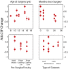
Potential contributions to visual improvements. The magnitude of contrast sensitivity development, via the change in area under the log contrast sensitivity function (AULCSF), is considered as a function of four factors: age at surgery, months since surgery, presurgery acuity (FC, finger counting at the specified distance; HM, hand movement), and the type of cataract (“mixed” indicates that the subject had different types of cataract in each eye). None of the four factors showed significant relationships (P > 0.05) with observed AULCSF changes.
Fig. 4.
Fig. 4.
Normal contrast sensitivity development. (Left) To summarize and compare previous studies of infant and childhood contrast sensitivity development, we normalized the AULCSF values reported for different ages to the earliest AULCSF obtained in each study: either 1–2 mo or 4 y old. For normal development, the change in AULCSF from 2 to 8 mo is approximately the same as from 4 y to maturity. (Right) For reference, the pattern of normal development can be used to predict how much AULCSF change is expected over 6 mo, as a function of age (black line). For the six patients who showed significant contrast sensitivity change in Fig. 1, AULCSF changes are plotted as a function of age at surgery (red dots). To compare with other demonstrations of adult visual plasticity, AULCSF changes due to video-game playing are presented (blue and green dots) (35, 36).
Fig. 5.
Fig. 5.
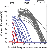
Survey of contrast sensitivity. A cross-sectional evaluation of Project Prakash patients compares contrast sensitivity assessed before surgery (red) and 6 mo to 5 y after surgery (blue). CSFs of age-matched normally sighted subjects are also shown (gray). Patients who did not have measurable CSFs at the time of testing are marked with an “x.” Contrast sensitivity development, and a general improvement in pattern vision, is evident with sight onset, although outcomes are variable across this unique sample.

References

    1. Wiesel TN, Hubel DH. Comparison of the effects of unilateral and bilateral eye closure on cortical unit responses in kittens. J Neurophysiol. 1965;28(6):1029–1040. - PubMed
    1. Movshon JA, et al. Effects of early unilateral blur on the macaque’s visual system. III. Physiological observations. J Neurosci. 1987;7(5):1340–1351. - PMC - PubMed
    1. Movshon JA, Kiorpes L. In: Early Visual Development, Normal and Abnormal. Simons K, editor. New York: Oxford Univ Press; 1993. pp. 296–305.
    1. Braastad BO, Heggelund P. Development of spatial receptive-field organization and orientation selectivity in kitten striate cortex. J Neurophysiol. 1985;53(5):1158–1178. - PubMed
    1. Daw NW, Wyatt HJ. Kittens reared in a unidirectional environment: Evidence for a critical period. J Physiol. 1976;257(1):155–170. - PMC - PubMed

Publication types