. 2014 Feb 4;111(5):1933-8.
doi: 10.1073/pnas.1323719111. Epub 2014 Jan 21.

Differential expression of estrogen receptor α, β1, and β2 in lobular and ductal breast cancer

Affiliations

Differential expression of estrogen receptor α, β1, and β2 in lobular and ductal breast cancer

Bo Huang et al. Proc Natl Acad Sci U S A. .

Abstract

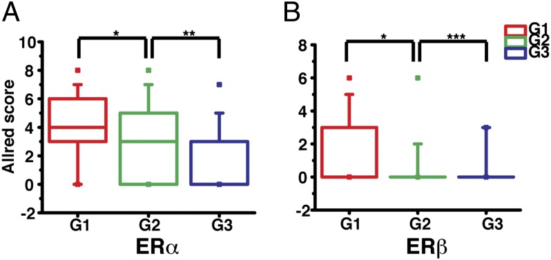
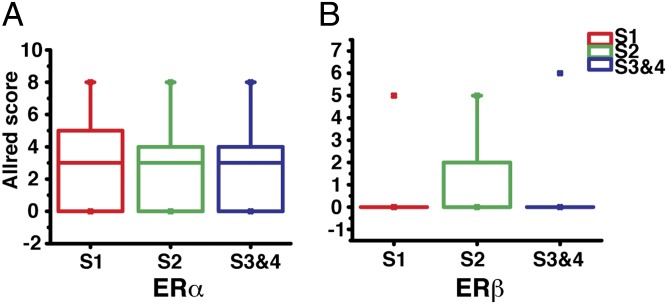
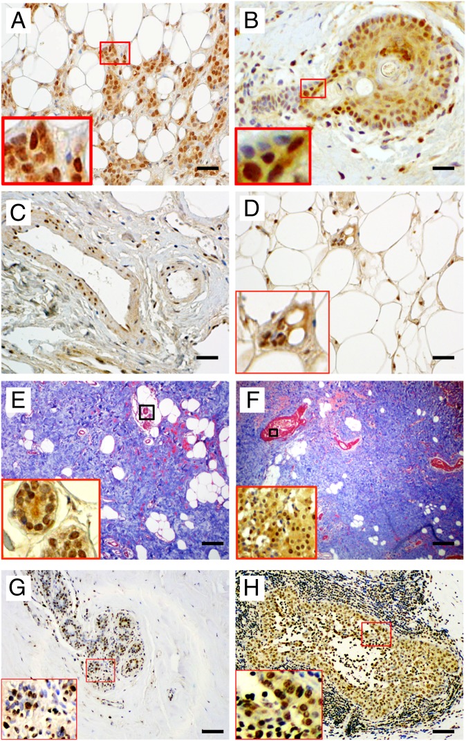
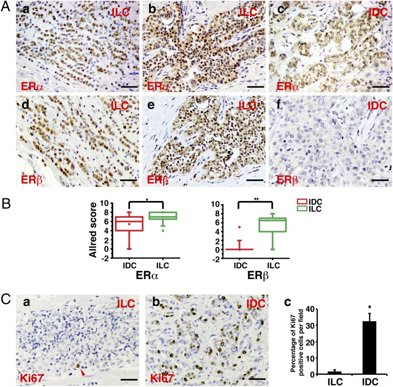
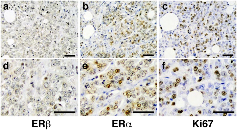
The role of estrogen receptor (ER) α as a target in treatment of breast cancer is clear, but those of ERβ1 and ERβ2 in the breast remain unclear. We have examined expression of all three receptors in surgically excised breast samples from two archives: (i): 187 invasive ductal breast cancer from a Japanese study; and (ii) 20 lobular and 24 ductal cancers from the Imperial College. Samples contained normal areas, areas of hyperplasia, and in situ and invasive cancer. In the normal areas, ERα was expressed in not more than 10% of epithelium, whereas approximately 80% of epithelial cells expressed ERβ. We found that whereas ductal cancer is a highly proliferative, ERα-positive, ERβ-negative disease, lobular cancer expresses both ERα and ERβ but with very few Ki67-positive cells. ERβ2 was expressed in 32% of the ductal cancers, of which 83% were postmenopausal. In all ERβ2-positive cancers the interductal space was filled with dense collagen, and cell nuclei expressed hypoxia-inducible factor 1α. ERβ2 expression was not confined to malignant cells but was strong in stromal, immune, and endothelial cells. In most of the high-grade invasive ductal cancers neither ERα nor ERβ was expressed, but in the high-grade lobular cancer ERβ was lost and ERα and Ki67 expression were abundant. The data show a clear difference in ER expression between lobular and ductal breast cancer and suggest (i) that tamoxifen may be more effective in late than in early lobular cancer and (ii) a potential role for ERβ agonists in preventing in situ ductal cancers from becoming invasive.

Keywords: ductal carcinoma in situ; invasive ductal carcinoma; invasive lobular carcinoma.


Conflict of interest statement

The authors declare no conflict of interest.

Figures

Fig. 1.
Fig. 1.
Expression of ERα and ERβ in normal tissue, DCIS, and IDC. (A) Representative immunohistochemical staining of ERα and ERβ in normal breast tissue, DCIS, and IDC. (Scale bars, 25 µm.) (B) Box chart of Allred scores of ERα and ERβ (n = 94) staining. Bottoms and tops of the boxes are the 25th and 75th percentiles, respectively; the lines across the boxes are the median values; the ends of the whiskers represent 10th and 90th percentiles; asterisks represent the minimum and maximum of all of the data (*P < 0.0001). (C) Box chart of Allred scores of ERα and ERβ staining (n = 115). Description of box chart is the same as in B (*P < 0.0001, **P = 0.01). (D) Box chart of Allred scores of ERα and ERβ (n = 94) staining. Description of the box chart is the same as in B (*P < 0.0001, **P = 0.03).
Fig. 2.
Fig. 2.
Correlation of ERα and ERβ expression with histological grades in IDC. The Allred scores of ERα (A) and ERβ (B) in IDC were sorted according to breast tumor histological grades (G1 to G3) and their expression differences between each grade were determined by statistical analysis (*P < 0.001, **P < 0.05, ***P = 0.61). The description of the box chart is the same as in Fig. 1B.
Fig. 3.
Fig. 3.
Correlation of ERα and ERβ expression with cancer stages in IDC. The Allred scores of ERα (A) and ERβ (B) in IDC were sorted according to breast cancer stages (S1, S2, and S3&4). Statistical analysis showed that there is no significant difference between each group. The description of the box chart is the same as in Fig. 1B.
Fig. 4.
Fig. 4.
ERβ2 expression in invasive breast cancers. (A–D and the Insets of E and F) Representative immunohistochemical staining of ERβ2 showing ERβ2 expression in cells from invading tumor edge (A); in cells surrounding necrotic centers (B); in stroma and in endothelium and muscle layers of the vessel wall (C); and in adipocytes (D). (E and F) Representative trichrome staining to show collagen in dense breasts (Insets are ERβ2 staining). (G and H) Representative HIF-1α staining. (Scale bars, 50 µm in A–D; 100 µm in E–H.)
Fig. 5.
Fig. 5.
ERα, ERβ, and Ki67 in IDC vs. ILC. (A) Representative immunohistochemical staining of ERα (ac) and ERβ (df) expression in ILC and IDC. (Scale bar, 50 μm.) (B) Box chart of Allred scores of ERα (Left) and ERβ (Right) staining. The description of the box chart is the same as in Fig. 1B (*P = 0.01, **P < 0.0001). (C) Comparison of Ki67 staining in IDC vs. ILC. Representative immunohistochemical staining of Ki67 in ILC and IDC is shown in a and b, respectively; c represents the quantitation analysis of the percentage of Ki67-positive cells (*P < 0.01).
Fig. 6.
Fig. 6.
Expression of (A and D) ERβ, (B and E) ERα, and (C and F) Ki67 in high-grade, advanced-stage ILC. (Scale bar, 50 µm.)

References

    1. Wellings SR, Jensen HM. On the origin and progression of ductal carcinoma in the human breast. J Natl Cancer Inst. 1973;50(5):1111–1118. - PubMed
    1. Platet N, Cathiard AM, Gleizes M, Garcia M. Estrogens and their receptors in breast cancer progression: A dual role in cancer proliferation and invasion. Crit Rev Oncol Hematol. 2004;51(1):55–67. - PubMed
    1. Warner M, Nilsson S, Gustafsson JA. The estrogen receptor family. Curr Opin Obstet Gynecol. 1999;11(3):249–254. - PubMed
    1. Osborne CK. Steroid hormone receptors in breast cancer management. Breast Cancer Res Treat. 1998;51(3):227–238. - PubMed
    1. Kuiper GG, Enmark E, Pelto-Huikko M, Nilsson S, Gustafsson JA. Cloning of a novel receptor expressed in rat prostate and ovary. Proc Natl Acad Sci USA. 1996;93(12):5925–5930. - PMC - PubMed

Publication types

MeSH terms

LinkOut - more resources