

Acoustic droplet vaporization is initiated by superharmonic focusing
- PMID: 24449879
- PMCID: PMC3918756
- DOI: 10.1073/pnas.1312171111
Acoustic droplet vaporization is initiated by superharmonic focusing
Abstract
Acoustically sensitive emulsion droplets composed of a liquid perfluorocarbon have the potential to be a highly efficient system for local drug delivery, embolotherapy, or for tumor imaging. The physical mechanisms underlying the acoustic activation of these phase-change emulsions into a bubbly dispersion, termed acoustic droplet vaporization, have not been well understood. The droplets have a very high activation threshold; its frequency dependence does not comply with homogeneous nucleation theory and localized nucleation spots have been observed. Here we show that acoustic droplet vaporization is initiated by a combination of two phenomena: highly nonlinear distortion of the acoustic wave before it hits the droplet and focusing of the distorted wave by the droplet itself. At high excitation pressures, nonlinear distortion causes significant superharmonics with wavelengths of the order of the droplet size. These superharmonics strongly contribute to the focusing effect; therefore, the proposed mechanism also explains the observed pressure thresholding effect. Our interpretation is validated with experimental data captured with an ultrahigh-speed camera on the positions of the nucleation spots, where we find excellent agreement with the theoretical prediction. Moreover, the presented mechanism explains the hitherto counterintuitive dependence of the nucleation threshold on the ultrasound frequency. The physical insight allows for the optimization of acoustic droplet vaporization for therapeutic applications, in particular with respect to the acoustic pressures required for activation, thereby minimizing the negative bioeffects associated with the use of high-intensity ultrasound.
Conflict of interest statement
The authors declare no conflict of interest.
Figures

 propagating from left to right, scattered wave
  outside the droplet and refracted wave
  inside the droplet.
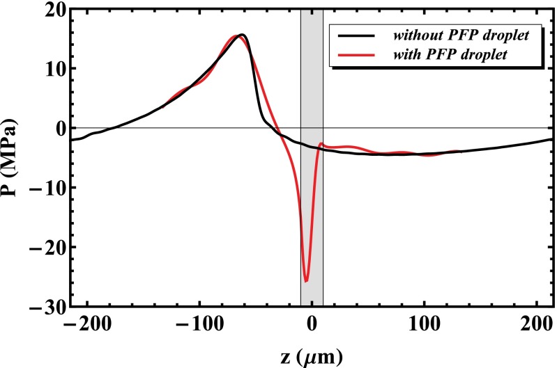
 and the pressure is amplified 5.8 times compared with the incident acoustic pressure
  .
.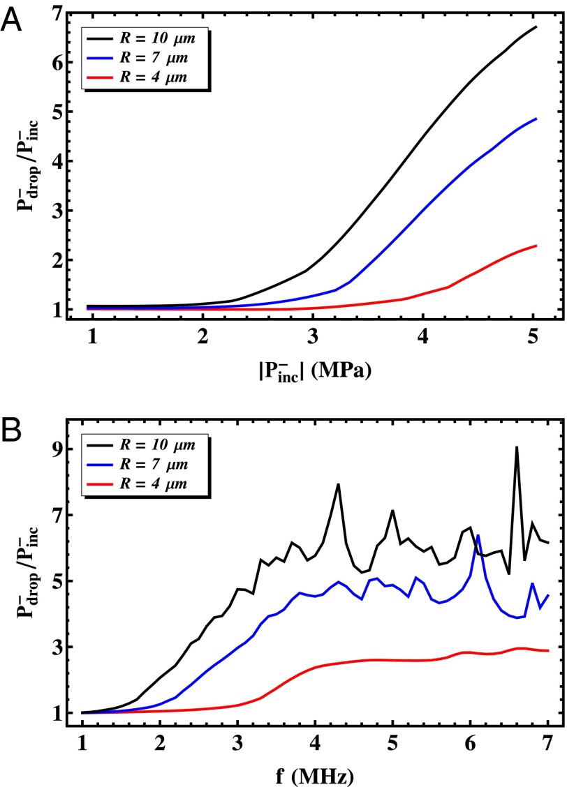
 at a driving frequency of 3.5 MHz and (B) as a function of the driving frequency f for a peak negative pressure of −4.5 MPa.
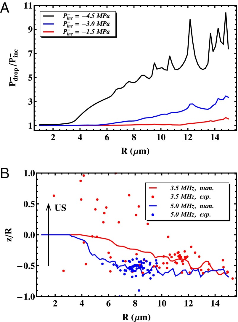
 . (B) The position of the nucleation site as a function of the droplet size. The position is taken along the axis of ultrasound propagation on the axis of symmetry of the system. The solid lines represent the calculated positions of maximal peak negative pressure for a frequency of 3.5 MHz (red) and 5.0 MHz (blue), respectively. The circles represent measured nucleation spots for a range of droplet sizes for the two frequencies.
References
-
- Duncan R. The dawning era of polymer therapeutics. Nat Rev Drug Discov. 2003;2(5):347–360. - PubMed
-
- Ferrari M. Cancer nanotechnology: Opportunities and challenges. Nat Rev Drug Discov. 2005;5(3):161–171. - PubMed
-
- Torchilin VP. Recent advances with liposomes as pharmaceutical carriers. Nat Rev Drug Discov. 2005;4(2):145–160. - PubMed
-
- Davis ME, Chen Z, Shin D. Nanoparticle therapeutics: An emerging treatment modality for cancer. Nat Rev Drug Discov. 2008;7(9):771–782. - PubMed
Publication types
MeSH terms
Substances
LinkOut - more resources
Full Text Sources
Other Literature Sources
Molecular Biology Databases
