. 2014 Feb 18;111(7):2728-33.
doi: 10.1073/pnas.1320755111. Epub 2014 Jan 21.

Structural basis for nuclear import of splicing factors by human Transportin 3

Affiliations

Structural basis for nuclear import of splicing factors by human Transportin 3

Goedele N Maertens et al. Proc Natl Acad Sci U S A. .

Abstract

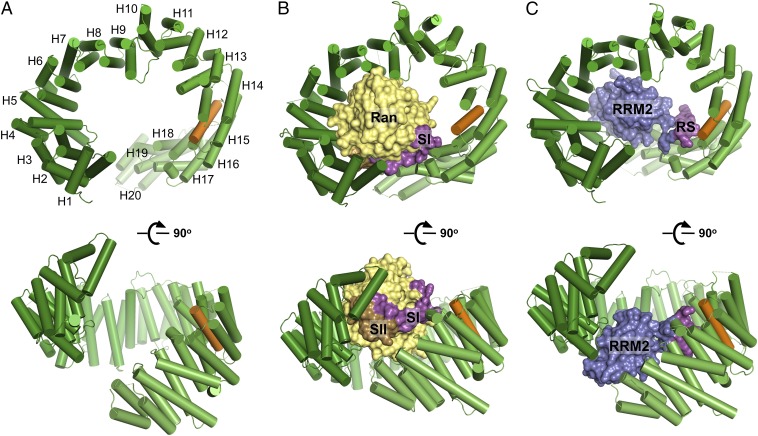
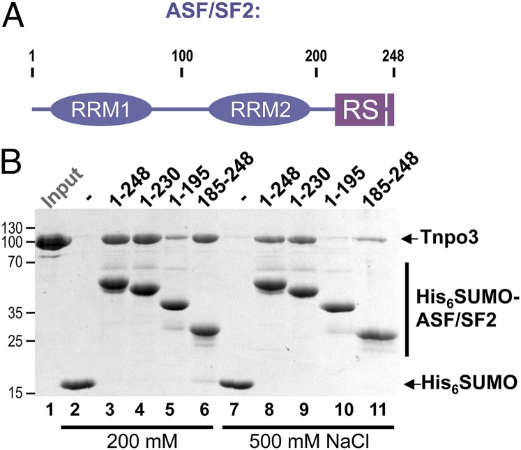
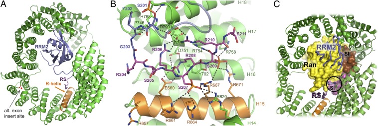
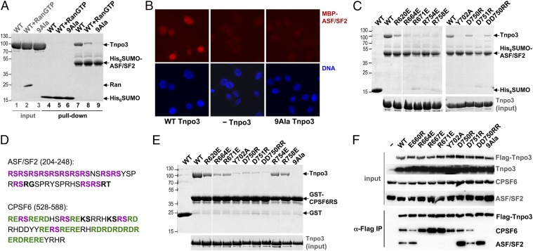
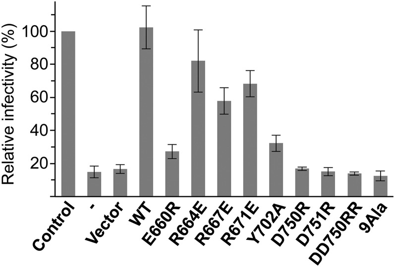
Transportin 3 (Tnpo3, Transportin-SR2) is implicated in nuclear import of splicing factors and HIV-1 replication. Herein, we show that the majority of cellular Tnpo3 binding partners contain arginine-serine (RS) repeat domains and present crystal structures of human Tnpo3 in its free as well as GTPase Ran- and alternative splicing factor/splicing factor 2 (ASF/SF2)-bound forms. The flexible β-karyopherin fold of Tnpo3 embraces the RNA recognition motif and RS domains of the cargo. A constellation of charged residues on and around the arginine-rich helix of Tnpo3 HEAT repeat 15 engage the phosphorylated RS domain and are critical for the recognition and nuclear import of ASF/SF2. Mutations in the same region of Tnpo3 impair its interaction with the cleavage and polyadenylation specificity factor 6 (CPSF6) and its ability to support HIV-1 replication. Steric incompatibility of the RS domain and RanGTP engagement by Tnpo3 provides the mechanism for cargo release in the nucleus. Our results elucidate the structural bases for nuclear import of splicing factors and the Tnpo3-CPSF6 nexus in HIV-1 biology.

Keywords: SR protein; Transportin-SR; host factor; importin.


Conflict of interest statement

The authors declare no conflict of interest.

Figures

Fig. 1.
Fig. 1.
Overview of the crystal structures of unliganded Tnpo3 (A), Tnpo3–RanGTP (B), and Tnpo3–ASF/SF2 (C) complexes. Tnpo3 is shown as cartoons, and ASF/SF2 and Ran are in space-fill mode. Tnpo3 is colored green, except the R-helix, which is orange; Switch I (SI) and Switch II (SII) regions of Ran are magenta and brown, respectively, and the remainder of the Ran structure is yellow. RRM2 and RS domains of ASF/SF2 are blue and magenta, respectively.
Fig. 2.
Fig. 2.
Interaction of ASF/SF2 with Tnpo3 in vitro. (A) Schematic of ASF/SF2 domain organization. (B) Pulldown of Tnpo3 with His6SUMO (lanes 2 and 7), full length His6SUMO–ASF/SF2 (lanes 3 and 8), or its deletion constructs (lanes 4–6 and 9–11) on Ni-nitrilotriacetic acid agarose in buffer containing 200 mM (lanes 2–6) or 500 mM (lanes 7–11) NaCl. The input quantity of Tnpo3 is shown in lane 1.
Fig. 3.
Fig. 3.
Details of the Tnpo3–ASF/SF2 structure. (A) Overview with both protein chains displayed as cartoons. The ASF/SF2 RRM2 and RS domains, the R-helix of Tnpo3, and the position of the insertion site of the alternative exon found in Transportin-SR (red asterisk) are indicated. (B) Details of the interface between Tnpo3 and the ASF/SF2 RS domain. (C) Superposition of the Tnpo3–RanGTP and Tnpo3–ASF/SF2 structures based on the HEAT repeats of the RS domain-binding region of Tnpo3. Ran, ASF/SF2, and Tnpo3 are shown in space-fill, Cα trace, and ribbon cartoon modes, respectively. The clash between Switch I of Ran and ASF/SF2 residues preceding the RS domain is indicated with a circle.
Fig. 4.
Fig. 4.
Mutagenesis of Tnpo3 residues involved in the interaction with the RS domain. (A) Pulldown of WT and 9Ala Tnpo3 with His6SUMO–ASF/SF2 in the presence and absence of RanGTP. (B) Nuclear import of fluorescently labeled MBP–ASF/SF2 in digitonin-permeabilized HeLa cells in the presence (Left) or absence (Center) of WT Tnpo3 or in the presence of the 9Ala Tnpo3 mutant (Right). The images show detection of MBP–ASF/SF2 (Upper) or DNA (Lower). (C) Pulldown of WT and mutant Tnpo3 proteins with His6SUMO–ASF/SF2. (D) Amino acid sequences of the C-terminal regions of human ASF/SF2 and CPSF6, spanning their RS domains. The canonical Arg-Ser dipeptides and the RS-like motifs Arg-Asp/Glu are in bold magenta and green, respectively, and RS-like Arg-Thr/Gly and Lys-Ser are in bold black. (E) Pulldown of WT and mutant Tnpo3 proteins with phosphorylated GST–CPSF6RS on glutathione beads. (F) Co-IP of endogenous ASF/SF2 and CPSF6 with WT and mutant Flag–Tnpo3 from cells depleted for expression of endogenous Tnpo3.
Fig. 5.
Fig. 5.
Effects of mutations within RS domain binding patch of Tnpo3 on HIV-1 infectivity. Relative HIV-1 infectivity measured in Tnpo3 knock-down cells before and after transduction with the empty vector, or vectors expressing WT or mutant Flag–Tnpo3 proteins, relative to that in control cell line (set as 100%). The error bars correspond to SDs from experiments done in triplicate.

References

    1. Stewart M. Molecular mechanism of the nuclear protein import cycle. Nat Rev Mol Cell Biol. 2007;8(3):195–208. - PubMed
    1. Görlich D, Panté N, Kutay U, Aebi U, Bischoff FR. Identification of different roles for RanGDP and RanGTP in nuclear protein import. EMBO J. 1996;15(20):5584–5594. - PMC - PubMed
    1. Strom AC, Weis K. Importin-beta-like nuclear transport receptors. Genome Biol. 2001;2(6):reviews 3008.1–3008.9. - PMC - PubMed
    1. Cook A, Bono F, Jinek M, Conti E. Structural biology of nucleocytoplasmic transport. Annu Rev Biochem. 2007;76:647–671. - PubMed
    1. Cook AG, Conti E. Nuclear export complexes in the frame. Curr Opin Struct Biol. 2010;20(2):247–252. - PubMed

Publication types

MeSH terms