Review
. 2014 Apr;24(3):205-20.
doi: 10.1111/bpa.12125. Epub 2014 Feb 25.

Schwannomas and their pathogenesis

Affiliations
Review

Schwannomas and their pathogenesis

David A Hilton et al. Brain Pathol. 2014 Apr.

Abstract

Schwannomas may occur spontaneously, or in the context of a familial tumor syndrome such as neurofibromatosis type 2 (NF2), schwannomatosis and Carney's complex. Schwannomas have a variety of morphological appearances, but they behave as World Health Organization (WHO) grade I tumors, and only very rarely undergo malignant transformation. Central to the pathogenesis of these tumors is loss of function of merlin, either by direct genetic change involving the NF2 gene on chromosome 22 or secondarily to merlin inactivation. The genetic pathways and morphological features of schwannomas associated with different genetic syndromes will be discussed. Merlin has multiple functions, including within the nucleus and at the cell membrane, and this review summarizes our current understanding of the mechanisms by which merlin loss is involved in schwannoma pathogenesis, highlighting potential areas for therapeutic intervention.

Keywords: Carney's complex; NF2; merlin; pathogenesis; schwannoma; schwannomatosis.


Figures

Figure 1
Figure 1
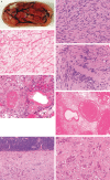
A. Well‐circumscribed lumbar region schwannoma, with attached nerve root. Characteristic histological features include cellular Antoni A tissue (B), with less cellular areas of loosely textured microcystic Antoni B tissue (C), and parallel arrays of nuclei forming a Verocay body (D). Blood vessels often show hyaline degeneration of their walls and thrombosis (E), and occasional areas of necrosis may be seen (F). A prominent pericapscular lymphocytic infiltrate is common in gastrointestinal schwannomas (G). Epitheloid morphology is seen in some benign schwannomas (H).
Figure 2
Figure 2
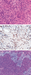
Cellular schwannomas are predominantly composed of Antoni A tissue and may show prominent nucleoli and mitotic activity (A). Like classical schwannomas, they show widespread S100 (B) and sometimes strong glial fibrillary acidic protein (C) immunoreactivity.
Figure 3
Figure 3
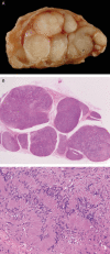
Plexiform schwannoma has a multinodular architecture seen macroscopically (A) and in histological sections (B). They are predominantly composed of Antoni A tissue and sometimes form Verocay bodies (C).
Figure 4
Figure 4
Melanotic schwannoma has prominent melanin pigment and nucleoli, often with intranuclear pseudoinclusions (A), prominent cytoplasmic vacuolation and rare calcified psammoma bodies (inset) (B). These tumors are immunoreactive with antibodies to melanosome proteins such as HMB‐45 (C) and basement membrane proteins such as laminin (D).
Figure 5
Figure 5
Whorl formation is seen more commonly in schwannomatosis and neurofibromatosis type 2 (NF2) (A). INI‐1 immunoreactivity often shows a mosaic pattern in familial schwannomas, including cases of NF2 (B). Alcian blue stain showing an area of myxoid change within a schwannoma from a patient with schwannomatosis (C).
Figure 6
Figure 6
Diagram summarizing genetic pathways leading to schwannoma pathogenesis.
Figure 7
Figure 7
Diagram illustrating key pathways dysregulated in merlin−/− tumor cells. Merlin loss leads to increased growth factor expression and activation of the Ras and phosphatidylinositol 3‐kinase (PI3K) pathways. A central mechanism is the loss of merlin‐induced inhibition of the CRL4–DCAF1 complex within the nucleus, resulting in increased transcription of a number of genes, including integrins and growth factor receptors. Merlin also interacts with cell surface proteins, including CD44 and adhesion junction proteins, so that merlin deficiency leads to reduced contact‐dependant cell cycle arrest. EGF = epidermal growth factor; ErbB2 = epidermal growth factor receptor 2; Gas 6 = growth arrest specific 6; IGFR = insulin‐like growth factor receptor; NRG = neuregulin; PDGFR = platelet‐derived growth factor receptor; VEGFR = vascular endothelial growth factor receptor.

References

    1. Agaram NP, Prakash S, Antonescu CR (2005) Deep‐seated plexiform schwannoma: a pathologic study of 16 cases and comparative analysis with the superficial variety. Am J Surg Pathol 29:1042–1048. - PubMed
    1. Ahmad Z, Brown CM, Patel AK, Ryan AF, Ongkeko R, Doherty JK (2010) Merlin knockdown in human Schwann cells: clues to vestibular schwannoma tumorigenesis. Otol Neurotol 31:460–466. - PMC - PubMed
    1. Akhmametyeva EM, Mihaylova MM, Luo H, Kharzai S, Welling DB, Chang LS (2006) Regulation of the neurofibromatosis 2 gene promoter expression during embryonic development. Dev Dyn 235:2771–2785. - PubMed
    1. Alfthan K, Heiska L, Grönholm M, Renkema GH, Carpén O (2004) Cyclic AMP‐dependent protein kinase phosphorylates merlin at serine 518 independently of p21‐activated kinase and promotes merlin‐ezrin heterodimerization. J Biol Chem 279:18559–18566. - PubMed
    1. Ammoun S, Cunliffe CH, Allen JC, Chiriboga L, Giancotti FG, Zagzag D et al (2010) ErbB/HER receptor activation and preclinical efficacy of lapatinib in vestibular schwannoma. Neuro Oncol 12:834–843. - PMC - PubMed