Review
. 2014 Jul 1;21(1):154-76.
doi: 10.1089/ars.2013.5773. Epub 2014 Mar 6.

Exercise-induced skeletal muscle remodeling and metabolic adaptation: redox signaling and role of autophagy

Affiliations
Review

Exercise-induced skeletal muscle remodeling and metabolic adaptation: redox signaling and role of autophagy

Elisabetta Ferraro et al. Antioxid Redox Signal. .

Abstract

Significance: Skeletal muscle is a highly plastic tissue. Exercise evokes signaling pathways that strongly modify myofiber metabolism and physiological and contractile properties of skeletal muscle. Regular physical activity is beneficial for health and is highly recommended for the prevention of several chronic conditions. In this review, we have focused our attention on the pathways that are known to mediate physical training-induced plasticity.

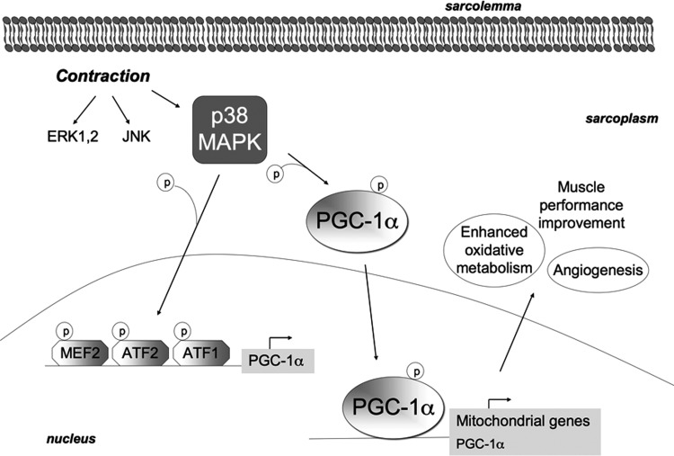
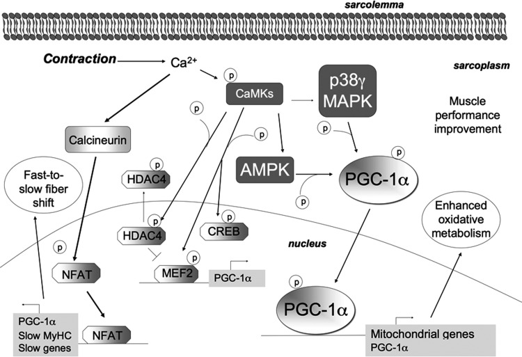
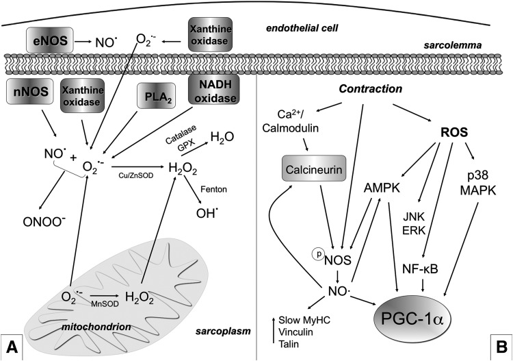
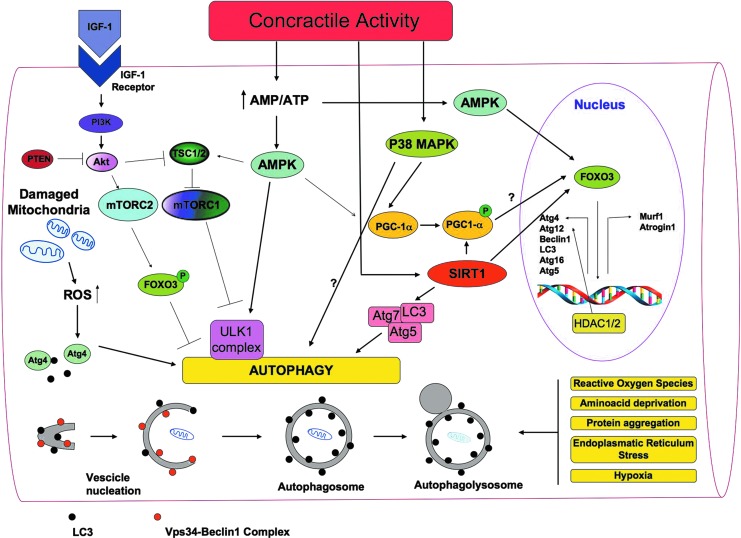
Recent advances: An important role for redox signaling has recently been proposed in exercise-mediated muscle remodeling and peroxisome proliferator-activated receptor γ (PPARγ) coactivator-1α (PGC-1α) activation. Still more currently, autophagy has also been found to be involved in metabolic adaptation to exercise.

Critical issues: Both redox signaling and autophagy are processes with ambivalent effects; they can be detrimental and beneficial, depending on their delicate balance. As such, understanding their role in the chain of events induced by exercise and leading to skeletal muscle remodeling is a very complicated matter. Moreover, the study of the signaling induced by exercise is made even more difficult by the fact that exercise can be performed with several different modalities, with this having different repercussions on adaptation.

Future directions: Unraveling the complexity of the molecular signaling triggered by exercise on skeletal muscle is crucial in order to define the therapeutic potentiality of physical training and to identify new pharmacological compounds that are able to reproduce some beneficial effects of exercise. In evaluating the effect of new "exercise mimetics," it will also be necessary to take into account the involvement of reactive oxygen species, reactive nitrogen species, and autophagy and their controversial effects.


Figures

<b>FIG. 1.</b>
FIG. 1.
Characteristics of mammalian skeletal muscle fiber types. The red color is associated with a high content of myoglobin. MyHC, myosin heavy-chain; SDH, succinate dehydrogenase; LDH, lactate dehydrogenase; CSA, cross-sectional area.
<b>FIG. 2.</b>
FIG. 2.
Metabolic pathways for ATP production in skeletal myofibers. (A) Skeletal muscles require a high amount of ATP for contraction. The main sources of energy are Glu and FFA. Glu uptake into the sarcoplasm from blood occurs, among other things, through the GLUT4. Once in the cytosol, Glu is phosphorylated by HK and forms Glu-6-P. One molecule of Glu-6-P can be converted into two molecules of Pyr through glycolysis, a metabolic anaerobic pathway involving 10 enzymes (the enzyme phosphofructokinase is an important control point in the glycolytic pathway). Depending on the energy needs, Glu-6-P can also be stored as glycogen. In anaerobic conditions Pyr is reduced to lactate by LDH. Alternatively, in aerobic conditions, Pyr might be transferred into the mitochondria matrix, where it is decarboxylated into acetyl-CoA by the PDH complex. Acetyl-CoA is then metabolized through the TCA cycle. The first enzyme acting in the TCA cycle is the citrate synthase that forms citrate from acetyl-CoA and oxaloacetate. The TCA cycle produces reducing equivalents (NADH, FADH2) and CO2. In addition to Pyr, another important source of acetyl-CoA is the β-oxidation of FFA. FFA enter the myofiber through a passive flip-flop or through a protein-mediated mechanism such as the FAT/CD36. In the cytosol, FFA undergo esterification and form triglycerides stored as lipid droplets that are surrounded by mitochondria. Alternatively, at the mitochondrial OM, they can be condensed with CoA to form FFA-CoA and, through the CPT1, they can cross the mitochondrial IM and reach the mitochondrial matrix where they undergo β-oxidation. β-oxidation is a cycle of four reactions. Each cycle produces a molecule of acetyl-CoA which, in turn, enters the TCA cycle. Along with acetyl-CoA, during β-oxidation, FADH2 and NADH are also formed. In skeletal muscles, at rest, excess of ATP produced is stored as PCr. ATP is converted into ADP and Pi by ATPase, and the Pi is used to convert Cr in PCr whose amount is roughly 10 times higher than the amount of ATP. During intense activity, PCr can anaerobically donate a phosphate group to ADP and form ATP for quick regeneration of ATP. PCr is, therefore, a rapid system to supply energy during contraction. The reversible phosphorylation of Cr is catalyzed by several CK. Once ATP also produced by PCr is consumed, the AK (myokinase) catalyzes the formation of ATP and AMP from two ADP molecules. During exercise, the amount of ATP produced by the myofiber increases enormously. However, the stores of ATP that can be detected in the myofiber are not as high, as ATP is stored in the form of PCr. (B) Reducing equivalents (NADH and FADH2) generated mainly during TCA, β-oxidation, and glycolysis are oxidized by the complexes of the respiratory chain (Complex I, II, III, and IV) in the oxidative phosphorylation pathway. Electrons are transferred from NADH and FADH2 to oxygen (which is reduced to H2O) by means of the enzyme complexes and by the electron carriers Ub and Cyt c of the respiratory chain. The energy released by reducing equivalent oxidation as electrons pass from one complex to the next is used to pump protons (H+) across the IM into the intermembrane space. This creates an electrochemical proton gradient across the IM, which is highly energetic. Protons can flow along this gradient through ATP synthase (ATPase or complex V); this backflow releases the energy of the proton gradient, which is used by ATP synthase to phosphorylate ADP and to form ATP. This phosphorylation of ADP is called oxidative, as it is coupled to the presence of oxygen that enables the oxidation of reducing equivalents. By this mechanism, nutrients are oxidated and their energy is stored in usable energy as ATP. ATP is also produced in a lower amount during glycolysis. OM, outer membrane; Glu, glucose; FFAs, free fatty acids; Glu-6-P, glucose-6-phosphate; Pyr, pyruvate; PDH, pyruvate dehydrogenase; TCA, tricarboxylic acid; FAT/CD36, fatty acyl translocase; CPT, carnitine palmitoyltransferase; CK, creatine kinases; AK, adenylate kinase; Ub, ubiquinon; IM, inner membrane; Cr, creatine; Cyt c, cytochrome c; GLUT4, glucose transporter 4; HK, hexokinase; PCr, phosphocreatine. To see this illustration in color, the reader is referred to the web version of this article at www.liebertpub.com/ars
<b>FIG. 3.</b>
FIG. 3.
Signaling pathways triggered by contraction and involving PGC-1α. PGC-1α is a major regulator of skeletal muscle remodeling induced by exercise. Changes in contractile activity are sensed by intracellular sensors involved in PGC-1α activation. These include Ca2+-dependent calcineurin and CAMKs, NO, ROS, p38 MAPK, AMPK, and SIRT1. Once activated, PGC-1α co-activates a variety of transcription factors and nuclear receptors such as PPARs, ERR-α, NRF-1, NRF-2, MEF2, CREB, thyroid receptor, FoxO, Sox9, and Tfam, which upregulate genes coding for mitochondrial proteins such as Mitofusin-2, PDK, Cyt c, and Cytrate synthase, thereby inducing mitochondrial biogenesis. PGC-1α also induces the transcription of genes encoding proteins that are involved in lipid metabolism (e.g., CPT, β-oxidation enzymes, and CD36), in angiogenesis, and in termogenesis. Moreover, PGC-1α triggers transcription of itself by interacting with MEF2 on its own promoter. By interacting with MEF2 (which binds the promoter of the gene encoding GLUT4), it also induces GLUT4 overexpression and enhances insulin sensitivity. The final effects of PGC-1α are fast-to-slow myofiber phenotype shift and muscle performance improvement. ERR-α, estrogen-related receptor-α; AMPK, AMP-activated protein kinase; CREB, cAMP-response element-binding protein; FoxO, forkhead box; Sox9, sex determining region Y-box 9; Tfam, transcription factor A mitochondrial; MEF2, myocyte enhancer factor 2; NRF=nuclear respiratory factor; p38 MAPK, p38 mitogen-activated protein kinase; PDK, pyruvate dehydrogenase kinase; PGC-1α=peroxisome proliferator-activated receptor γ (PPARγ) coactivator-1α; ROS, reactive oxygen species; SIRT1, sirtuin 1.
<b>FIG. 4.</b>
FIG. 4.
Modulation of PGC-1α by AMPK and SIRT1. The serine/threonine kinase AMPK is activated by increased AMP/ATP ratio. AMPK is a heterotrimer with a catalytic α subunit and two (β and γ) regulatory subunits. Exercise requires a high amount of ATP, thereby leading to AMP/ATP ratio increase, which enhances AMPK phosphorylation and AMPK enzymatic activity. AMPK acts by phosphorylating and modulating some transcription factors such as NRF-1, CREB, and MEF2, as well as HDACs, and also by phosphorylating metabolic enzymes. For example, it induces lipid metabolism by phosphorylating and inactivating ACC. In the skeletal muscle, AMPK activation also triggers Glu uptake by enhancing GLUT4 translocation to the sarcolemma. Moreover, AMPK phosphorylates PGC-1α; this is needed for its deacetylation by SIRT1. These modifications enable PGC-1α to migrate into the nucleus, where it plays its role as a transcription factor, thereby triggering numerous effects. ACC, acetyl-CoA carboxylase; HDAC, histone deacetylase.
<b>FIG. 5.</b>
FIG. 5.
MAPKs are activated by exercise. During acute exercise, three MAPKs are activated: specifically, ERK1/2, JNK, and p38 MAPK. p38 MAPK is able to phosphorylate PGC-1α and to favor its translocation to the nucleus, where it acts as a transcription factor for mitochondrial genes as well as for itself. In addition, p38 MAPK phosphorylates and activates the transcription factors MEF2, ATF1, and ATF2, which bind to the PGC-1α promoter, thereby inducing up-regulation of PGC-1α and, in turn, of mitochondrial genes. It has also been proposed that p38 MAPK might regulate PGC-1α induction by endurance exercise mainly by mediating its nuclear translocation; while PGC-1α up-regulation might occur as a secondary effect of PGC-1α translocation and activity. The increased activity of PGC-1α-induced by p38 MAPK mainly mediates angiogenesis and metabolic adaptation to exercise. ATF, activating transcription factor; ERK, extracellular-signal-regulated kinase; JNK, c-Jun N-terminal kinase.
<b>FIG. 6.</b>
FIG. 6.
Exercise adaptation is mediated by Ca2+ signaling. During exercise, Ca2+ concentration increases inside the myofiber and activates CaMKs. Calcineurin is a phosphatase that primarily dephosphorylates and activates NFAT. NFAT mainly promotes the transcription of PGC-1α and of slow genes (such as MyHC), thus most probably mediating the fast-to-slow myofiber transition. CaMKII is the main isoform of CaMKs in human skeletal muscle. On Ca2+ increase due to exercise, CaMKs become phosphorylated in an intensity-dependent manner; they, along with p38 MAPK, target PGC-1α and mitochondrial biogenesis. In addition, it is considered that CaMKs might also phosphorylate CREB, AMPK, and HDACs. CaMKs phosphorylate and directly activate MEF2, which promotes the transcription of PGC-1α. Nuclear HDAC4 inhibits MEF2. By phosphorylating HDAC4, CaMKs induce HDAC4 export out of the nucleus, thereby releasing MEF2. CaMKs, Ca2+/calmodulin-dependent kinases; NFAT, nuclear factor of activated T-cells.
<b>FIG. 7.</b>
FIG. 7.
ROS and RNS production and their role in exercise-mediated skeletal muscle plasticity. (A) Several ROS and RNS are produced in skeletal muscle both at rest and during contraction. NO is produced by NOS (eNOS in the endothelial cells and nNOS in the muscle fibers). The O2•− is generated into mitochondria in at least five sites (three of which being well characterized) within the mitochondrial respiratory chain through incomplete reduction of oxygen in the electron transport system. O2•− can also be generated as a specific product of some enzymes into mitochondria. Moreover, NADH oxidase, xanthine oxidase, and PLA2 are other O2•− generators. O2•− can undergo spontaneous dismutation or dismutation catalyzed by MnSOD (in the matrix) and Cu/ZnSOD (in the intermembrane space and in the cytosol) and form H2O2. H2O2 is cytotoxic; however, it is poorly reactive and is considered a relatively weak oxidizing agent. H2O2 is unable to oxidize DNA or lipids directly, but it can inactivate some enzymes. H2O2 might be de-tossificated in H2O by catalase or GPX or be transformed, through the Fenton reaction, into OH which is, by contrast, highly reactive and able to damage most macromolecules, including DNA, proteins, and lipids. O2•− can react with NO and produce ONOO. ONOO is able to rapidly cross membranes and is a strong oxidant agent that can lead to DNA damage and nitration of proteins. Sub-intracellular measurements and detection of ROS are usually prone to artifacts, while O2•− intracellular catabolism is extremely complex and controversial. Therefore, the earlier description is an oversimplification. Skeletal muscle has a well-developed system that prevents potentially deleterious effects of ROS (such as catalases, SODs, glutathione, GPX, peroxiredoxins, and thioredoxins). The abundance of scavengers abrogates free-radical chain reaction propagation under physiological conditions. If redox homeostasis is disrupted, the cell becomes damaged, thus leading to a pathological condition. (B) ROS and NO mediate a contraction-induced adaptive response to exercise. NO and ROS mediate the up-regulation of PGC-1α, GLUT4, mitochondrial genes, and slow genes. Moreover, ROS increase Glu uptake by triggering p38 MAPK (and also ERK) phosphorylation and activation, which possibly phosphorylates and activates PGC-1α. ROS and RNS trigger PGC-1α phosphorylation and Glu uptake also through AMPK. Moreover, ROS induce NF-κB-mediated transcription of PGC-α. Contraction also induces NO production by NOS. NO, in turn, activates calcineurin/NFAT induction of fast-to-slow phenotype. NOS is a Ca2+/calmodulin-dependent enzyme that should be dephosphorylated in order to produce NO, and this might depend on calcineurin. NOS is also regulated by AMPK. NO, nitric oxide; O2•−, superoxide anion; H2O2, hydrogen peroxide; GPX, glutatione peroxidase; NF-κB, nuclear factor-kappa B; NOS, nitric oxide synthases; OH, hydroxyl radical; ONOO, peroxynitrite; PLA2, phospholipase A2; RNS, reactive nitrogen species; SOD, superoxide dismutase.
<b>FIG. 8.</b>
FIG. 8.
A general overview of the signaling molecules involved in the regulation of autophagy in skeletal muscles. Autophagy is a multi-step process that involves distinct phases during which part of the cytoplasm intracellular organelles are sequestered within characteristic double-membraned autophagic vacuoles (autophagosomes) that fuse with lysosomes and become autophagolysosomes. There, defective intracellular organelles and proteins are digested by a battery of lysosomal hydrolases. LC3 and the Vps34-Beclin1 complex are required, among many other proteins, for autophagosome formation. In skeletal muscles, ROS production influences different cell signaling pathways, including selective mitochondrial autophagy (mitophagy). Damaged mitochondria removal is particularly needed during exercise when the oxidative metabolism and the turnover of mitochondria increase. Moreover, ROS are mainly produced by mitochondria. The activation of the pivotal autophagy protein LC3 is mediated by the redox-sensitive Atg4 protease, which cleaves LC3. Growth factors such as IGF induce the PI3K/Akt signaling, which activates mTORC1 and mTORC2. mTORC1 (known to activate protein synthesis) also inhibits autophagy, as it inhibits the formation of the Atg1 (in humans ULK1) complex. ULK1 is a serine/threonine kinase that forms a complex with different regulatory proteins such as Atg13 and Atg17. Atg13 hyper-phosphorylation inhibits its association with Atg1, while the Atg1–Atg13 interaction enables the generation of autophagosomes. mTORC2 phosphorylates and inhibits FoxO transcription factors, thus inhibiting autophagy. Indeed, dephosphorylated FoxOs migrate into the nucleus and activate the transcription of genes that control muscle mass. FoxOs activate both ubiquitin-proteasome genes (atrogin-1 and MuRF-1) and autophagy-lysosome genes such as Beclin1, LC3, Atg4, Atg12, Atg16, and Atg5. PTEN inhibits the PI3K/Akt signaling pathway, and, therefore, it enables autophagy. Exercise, mitochondrial dysfunctions, starvation, and oxidative stress increase the intracellular AMP/ATP ratio, thus activating the energy stress sensor AMPK, which, in turn, promotes autophagy by inhibiting mTORC1 through the phosphorylation of TSC2. The TSC complex, consisting of TSC1 and TSC2 proteins, regulates the activity of the mTORC via Rheb, a small GTPase. In addition, AMPK induces autophagy by triggering ULK1 phosphorylation. By integrating signals from upstream sensors such as mTOR and AMPK, the ULK1 complex plays a central role in autophagy. AMPK activates FoxO transcription factors and leads to the expression of LC3 and Beclin1. Through a mechanism not yet known, p38 MAPK also seems to induce autophagy. In addition, SIRT1 can deacetylate Atg5, Atg7, and LC3, thus inducing autophagy; nuclear SIRT1 might induce the expression of autophagy genes through the activation of FoxOs. HDAC1 and 2 regulate muscle autophagy by controlling the expression of autophagy genes. During exercise, PGC-1α induction is mediated, among other things, by AMPK and p38 MAPK. Exercise up-regulates SIRT1 that removes acetyl groups from PGC-1α, enabling its translocation to the nucleus. PGC-1α induction has been associated with increased autophagy, although this hypothesis needs further investigation. IGF, insulin growth factor; LC3, microtubule-associated protein 1-light chain 3; mTOR, mammalian target of rapamycin; mTORC, mTOR complex; MuRF-1, muscle ring finger protein 1; PI3K, phosphoinositide 3-kinase; PTEN, phosphatase and tensin homologue; TSC, tuberosus sclerosis complex; ULK1, unc-51-like kinase1; Vps34, vacuolar protein sorting 34. To see this illustration in color, the reader is referred to the web version of this article at www.liebertpub.com/ars

References

    1. Abruzzo PM, Esposito F, Marchionni C, di Tullio S, Belia S, Fulle S, Veicsteinas A, and Marini M. Moderate exercise training induces ROS-related adaptations to skeletal muscles. Int J Sports Med 34: 676–687, 2013 - PubMed
    1. Akimoto T, Pohnert SC, Li P, Zhang M, Gumbs C, Rosenberg PB, Williams RS, and Yan Z. Exercise stimulates Pgc-1alpha transcription in skeletal muscle through activation of the p38 MAPK pathway. J Biol Chem 280: 19587–19593, 2005 - PubMed
    1. Arico S, Petiot A, Bauvy C, Dubbelhuis PF, Meijer AJ, Codogno P, and Ogier-Denis E. The tumor suppressor PTEN positively regulates macroautophagy by inhibiting the phosphatidylinositol 3-kinase/protein kinase B pathway. J Biol Chem 276: 35243–35246, 2001 - PubMed
    1. Atherton PJ, Babraj J, Smith K, Singh J, Rennie MJ, and Wackerhage H. Selective activation of AMPK-PGC-1alpha or PKB-TSC2-mTOR signaling can explain specific adaptive responses to endurance or resistance training-like electrical muscle stimulation. FASEB J 19: 786–788, 2005 - PubMed
    1. Baar K, Wende AR, Jones TE, Marison M, Nolte LA, Chen M, Kelly DP, and Holloszy JO. Adaptations of skeletal muscle to exercise: rapid increase in the transcriptional coactivator PGC-1. FASEB J 16: 1879–1886, 2002 - PubMed

Publication types