. 2014 Jan 21;12(1):491-507.
doi: 10.3390/md12010491.

Hyaluromycin, a new hyaluronidase inhibitor of polyketide origin from marine Streptomyces sp

Affiliations

Hyaluromycin, a new hyaluronidase inhibitor of polyketide origin from marine Streptomyces sp

Enjuro Harunari et al. Mar Drugs. .

Abstract

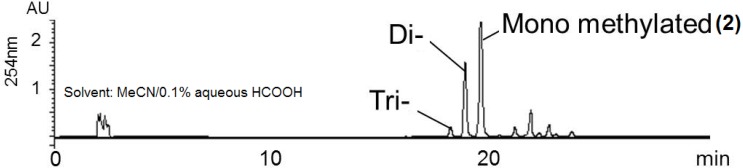
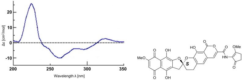
Hyaluromycin (1), a new member of the rubromycin family of antibiotics, was isolated from the culture extract of a marine-derived Streptomyces sp. as a HAase inhibitor on the basis of HAase activity screening. The structure of 1 was elucidated through the interpretation of NMR data for the compound and its 3″-O-methyl derivative in combination with an incorporation experiment with [1,2-13C2]acetate. The compound's absolute configuration was determined by the comparison of its circular dichroism (CD) spectrum with those of other rubromycins. Hyaluromycin (1) consists of a γ-rubromycin core structure possessing a 2-amino-3-hydroxycyclopent-2-enone (C5N) unit as an amide substituent of the carboxyl function; both structural units have been reported only from actinomycetes. Hyaluromycin (1) displayed approximately 25-fold more potent hyaluronidase inhibitory activity against hyaluronidase than did glycyrrhizin, a known inhibitor of plant origin.


Figures

Figure 1
Figure 1
Natural rubromycins.
Figure 2
Figure 2
Natural products containing the C5N substructure.
Figure 3
Figure 3
Families of compounds containing the C5N substructure.
Figure 4
Figure 4
Structure of hyaluromycin (1).
Figure 5
Figure 5
1H-1H COSY and HMBC correlations of compound 1.
Figure 6
Figure 6
HPLC chromatogram of methylated derivatives of 1.
Figure 7
Figure 7
13C NMR spectrum of [1,2-13C2]acetate-labeled 2.
Figure 8
Figure 8
1H-1H COSY and HMBC correlations of [1,2-13C2]acetate-labeled 2.
Figure 9
Figure 9
13C-13C couplings observed in 2D INADEQUATE (a,b) and 13C (c) NMR spectra of [1,2-13C2]acetate-labeled 2. The coupling of C-1″/C-5″ and C-3″/C-4″ were only observed in the 13C NMR spectrum (c). (a) Optimized for 1Jcc = 50 Hz; (b) Optimized for 1Jcc = 35 Hz; (c) 13C NMR spectra; (d) Observed in 2D INADEQUATE.
Figure 10
Figure 10
CD spectrum and absolute configuration of 2.

References

    1. Ohya T., Kaneko Y. Novel hyaluronidase from streptomyces. Biochim. Biophys. Acta. 1970;198:607–609. doi: 10.1016/0005-2744(70)90139-7. - DOI - PubMed
    1. Hamai A., Morikawa K., Horei K., Tokuyasu K. Purification and characterization of hyaluronidase from Streptococcus dysgalactiae. Agric. Biol. Chem. 1989;53:2163–2168. doi: 10.1271/bbb1961.53.2163. - DOI
    1. Baker J.R., Dong S., Pritchard D.G. The hyaluronan lyase of Streptococcus pyogenes bacteriophage H4489A. Biochem. J. 2002;365:317–322. doi: 10.1042/BJ20020149. - DOI - PMC - PubMed
    1. Kemeny D.M., Dalton N., Lawrence A.J., Pearce F.L., Vernon C.A. The purification and characterisation of hyaluronidase from the venom of the honey bee, Apis mellifera. Eur. J. Biochem. 1984;139:217–223. doi: 10.1111/j.1432-1033.1984.tb07997.x. - DOI - PubMed
    1. Kolarich D., Léonard R., Hemmer W., Altmann F. The N-glycans of yellow jacket venom hyaluronidases and the protein sequence of its major isoform in Vespula vulgaris. FEBS J. 2005;272:5182–5190. doi: 10.1111/j.1742-4658.2005.04841.x. - DOI - PubMed

MeSH terms