. 2013 Dec 23;1(1):31.
doi: 10.1186/2049-2618-1-31.

Community differentiation of the cutaneous microbiota in psoriasis

Affiliations

Community differentiation of the cutaneous microbiota in psoriasis

Alexander V Alekseyenko et al. Microbiome. .

Abstract

Background: Psoriasis is a common chronic inflammatory disease of the skin. We sought to characterize and compare the cutaneous microbiota of psoriatic lesions (lesion group), unaffected contralateral skin from psoriatic patients (unaffected group), and similar skin loci in matched healthy controls (control group) in order to discern patterns that govern skin colonization and their relationship to clinical diagnosis.

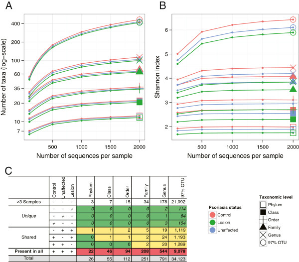
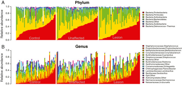
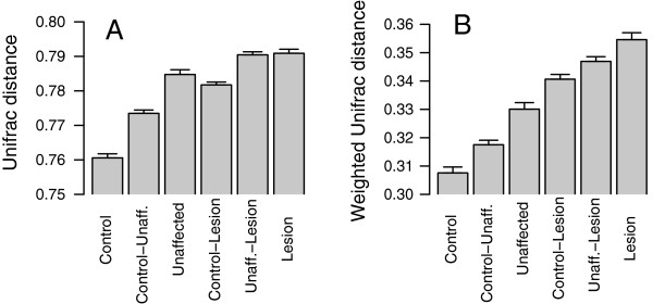
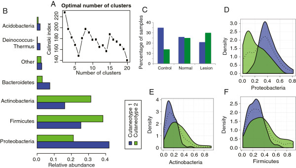
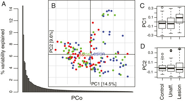
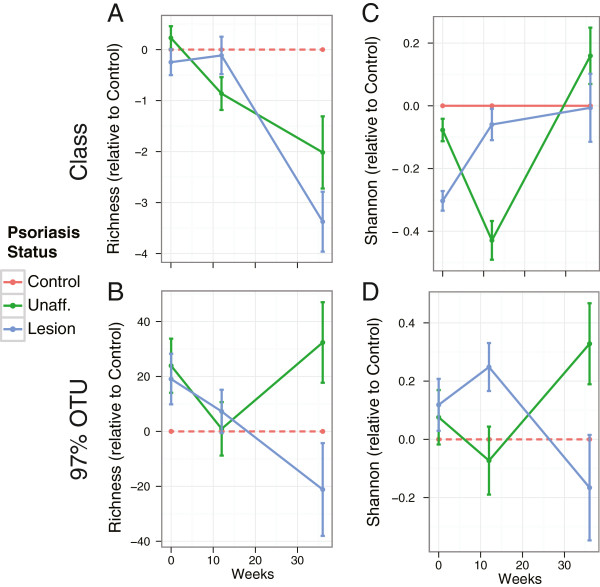
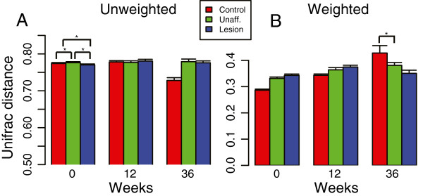
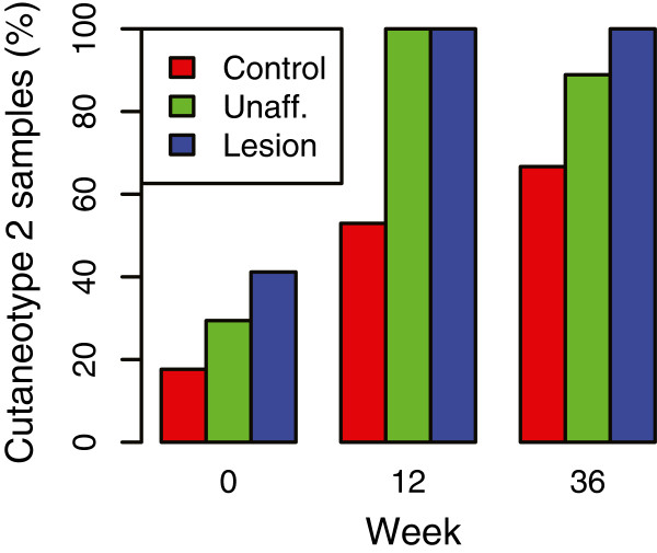
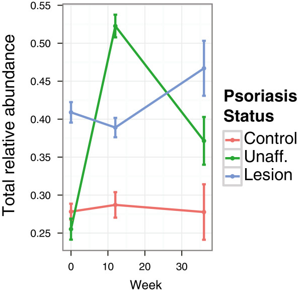
Results: Using high-throughput 16S rRNA gene sequencing, we assayed the cutaneous bacterial communities of 51 matched triplets and characterized these samples using community data analysis techniques. Intragroup Unifrac β diversity revealed increasing diversity from control to unaffected to lesion specimens. Likewise, principal coordinates analysis (PCoA) revealed separation of the lesion samples from unaffected and control along the first axis, suggesting that psoriasis is a major contributor to the observed diversity. The taxonomic richness and evenness decreased in both lesion and unaffected communities compared to control. These differences are explained by the combined increased abundance of the four major skin-associated genera (Corynebacterium, Propionibacterium, Staphylococcus, and Streptococcus), which present a potentially useful predictor for clinical skin type. Psoriasis samples also showed significant univariate decreases in relative abundances and strong classification performance of Cupriavidus, Flavisolibacter, Methylobacterium, and Schlegelella genera versus controls. The cutaneous microbiota separated into two distinct clusters, which we call cutaneotypes: (1) Proteobacteria-associated microbiota, and (2) Firmicutes-associated and Actinobacteria-associated microbiota. Cutaneotype 2 is enriched in lesion specimens compared to control (odds ratio 3.52 (95% CI 1.44 to 8.98), P <0.01).

Conclusions: Our results indicate that psoriasis induces physiological changes both at the lesion site and at the systemic level, which select for specific differential microbiota among the assayed clinical skin types. These differences in microbial community structure in psoriasis patients are potentially of pathophysiologic and diagnostic significance.


Figures

Figure 1
Figure 1
α Diversity rarefaction curves of cutaneous microbiota in psoriasis (lesion), unaffected and control specimens. (A) Taxonomical richness trends towards decreasing α diversity in unaffected and lesion specimens relative to control, with no statistically significant differences between skin types. (B) Shannon index is significantly different (decreases from control to unaffected to lesion) among skin types at all taxonomic levels (P <0.05), except at the operational taxonomical unit (OTU) level. (C) Analysis of taxa sharing. Taxa present in <3 samples excluded from the analysis. Taxa that are only observed in one clinical skin type are denoted as ‘unique’. Taxa that are present in two types of skin are denoted as ‘shared’. The data show that nearly all taxa are represented in all three types of skin. The shading represents the relative distribution (heatmap) for each column number (green = low, yellow = intermediate, red = high).
Figure 2
Figure 2
Taxonomical composition of cutaneous microbiota shown by skyline plots. (A) phylum, (B) genus levels. Predominant taxa are shown. White areas reflect specific taxa not graphed.
Figure 3
Figure 3
Intergroup and intragroup β diversity. Mean (± SEM) pairwise Unifrac distances obtained by unweighted (A) or weighted (B) methods are shown. Significance was determined by one-way analysis of variance (ANOVA) with the Tukey honestly significant difference (HSD) method for correction for multiple comparisons. For the 51 triplet sets, all differences in the unweighted Unifrac comparisons were significant (P <0.05), except for the comparisons between control-lesion and within unaffected, and the comparison between unaffected-lesion and within lesion. All differences between the weighted pairwise comparisons were significant (P <0.05) except for the comparisons between control-lesion and unaffected-lesion, and the comparison between unaffected-lesion and within lesion.
Figure 4
Figure 4
Evidence for psoriasis-associated cutaneotypes. (A) Clustering analysis of the Jensen-Shannon divergence distances indicated that two clusters, which we called cutaneotypes, can optimally describe the data. The Calinski-Harabasz index plot versus the number of clusters was used to select the optimal number of clusters, which also indicated that two has the highest Calinski index. (B) The relative abundance of major phyla is different between the two cutaneotypes. (C) The distribution of specimens in the three clinical categories is significantly associated with cutaneotype (P <0.01 based on χ2 test). Cutaneotype 2 is enriched for lesion specimens, while controls are more likely to be of cutaneotype 1 (OR 3.52, 95% CI 1.44 to 8.98), unaffected skin specimens are approximately evenly distributed between the cutaneotypes. Cutaneotype 1 is dominated by Proteobacteria(D). In the density plots, the dotted line shows the combined density for both cutaneotypes: blue, cutaneotype 1; green, cutaneotype 2. In cutaneotype 2, Actinobacteria(E) and Firmicutes(F) are the dominant phyla.
Figure 5
Figure 5
Principal coordinates analysis (PCoA) of weighted Unifrac distances. Based on separation of the relative values of the percentage variability explained by the principal axes, we chose to represent the cutaneous microbiome of our study in the first two components (A). (B) On the scatterplot of the first two principal axes of the PCoA, each point represents an individual subject (red = control, green = unaffected, blue = lesion) and the colored boxes are positioned at the geometrical center of all the points in the corresponding groups. The distributions of the points along PC1 (C) and PC2 (D) are shown in the boxes (representing the middle 50% of the data) and whiskers (95% interval) plots. Along PC1, lesion samples are significantly different from control (P <0.001) and unaffected (P <0.015) based on one-way analysis of variance (ANOVA) with Tukey honestly significant difference (HSD) multiple comparison correction.
Figure 6
Figure 6
α Diversity trends in the longitudinal cohort. For the longitudinal cohort, we show the α diversity ((A,B): richness, (C,D) Shannon) of unaffected (green) and lesion (blue) samples relative to the control group at the class level (A,C) and for 97% operational taxonomical units (OTUs) (B,D). At all timepoints, the data show no statistically significant intergroup differences in α diversity.
Figure 7
Figure 7
Intragroup β diversity trends in the longitudinal cohort. (A) Unweighted, and (B) weighted intragroup Unifrac distances. We show β diversity at three timepoints in relation to treatment of the patients with psoriasis, at 0 (pretreatment), 12, and 36 weeks of therapy. An asterisk (*) indicates comparisons that are significant at the 5% α level.
Figure 8
Figure 8
Distribution of the longitudinal samples in cutaneotype 2. The cutaneotype prevalence in the longitudinal cohort reflects that of the cross-sectional study (Figure 5C). Cutaneotype 2 is most prevalent in the unaffected and lesion specimens. The frequency of cutaneotype 2 is significantly different (P <0.005, Fisher exact test) at week 12 in control vs lesion and in control vs unaffected specimens. Other comparisons are not statistically significant.
Figure 9
Figure 9
Relative abundance of skin-associated genera in longitudinal samples. The combined abundance (± SEM) of four skin-associated genera (Corynebacterium, Propionibacterium, Staphylococcus and Streptococcus) is shown over the course of the longitudinal study in three skin types. Control skin demonstrates a stable relative abundance of these bacteria, while variability is evident in unaffected and lesion skin. Lesional microbiota shows a dramatic increase in these bacteria at all timepoints.

Similar articles

Cited by

References

    1. Nestle FO, Kaplan DH, Barker J. Psoriasis. N Engl J Med. 2009;361:496–509. doi: 10.1056/NEJMra0804595. - DOI - PubMed
    1. Fahlen A, Engstrand L, Baker BS, Powles A, Fry L. Comparison of bacterial microbiota in skin biopsies from normal and psoriatic skin. Arch Dermatol Res. 2012;304:15–22. doi: 10.1007/s00403-011-1189-x. - DOI - PubMed
    1. Paulino LC, Tseng CH, Strober BE, Blaser MJ. Molecular analysis of fungal microbiota in samples from healthy human skin and psoriatic lesions. J Clin Microbiol. 2006;44:2933–2941. doi: 10.1128/JCM.00785-06. - DOI - PMC - PubMed
    1. Paulino LC, Tseng CH, Blaser MJ. Analysis of Malassezia microbiota in healthy superficial human skin and in psoriatic lesions by multiplex real-time PCR. FEMS Yeast Res. 2008;8:460–471. doi: 10.1111/j.1567-1364.2008.00359.x. - DOI - PubMed
    1. Gao Z, Tseng CH, Strober BE, Pei Z, Blaser MJ. Substantial alterations of the cutaneous bacterial biota in psoriatic lesions. PLoS One. 2008;3:e2719. doi: 10.1371/journal.pone.0002719. - DOI - PMC - PubMed