. 2014 Apr;38(4):480-93.
doi: 10.1097/PAS.0000000000000165.

Whipple made simple for surgical pathologists: orientation, dissection, and sampling of pancreaticoduodenectomy specimens for a more practical and accurate evaluation of pancreatic, distal common bile duct, and ampullary tumors

Affiliations

Whipple made simple for surgical pathologists: orientation, dissection, and sampling of pancreaticoduodenectomy specimens for a more practical and accurate evaluation of pancreatic, distal common bile duct, and ampullary tumors

N Volkan Adsay et al. Am J Surg Pathol. 2014 Apr.

Abstract

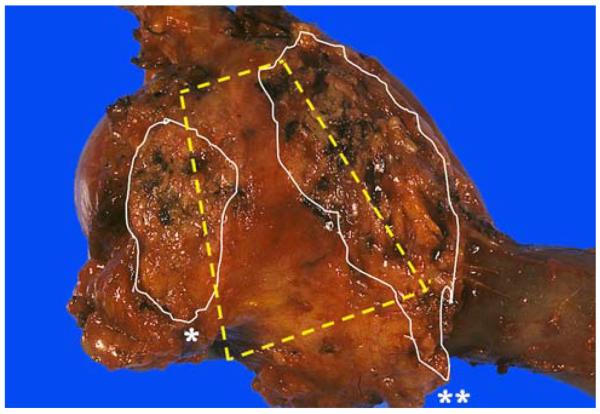
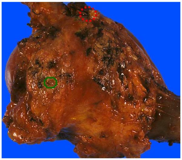
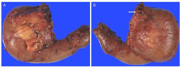
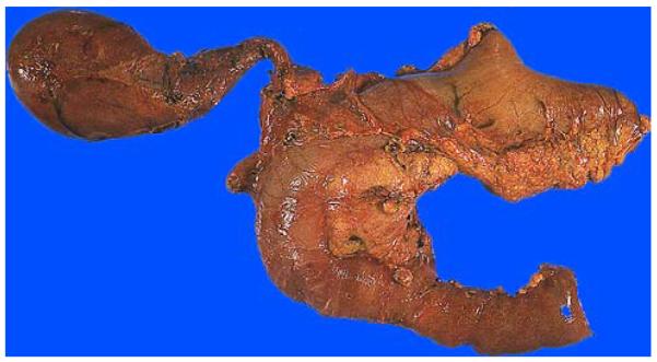
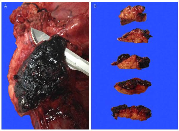
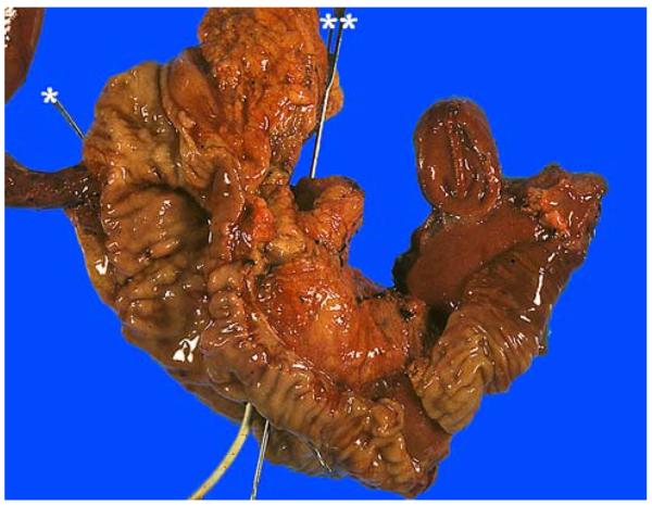
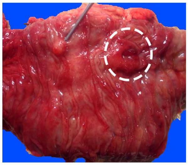
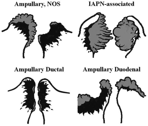
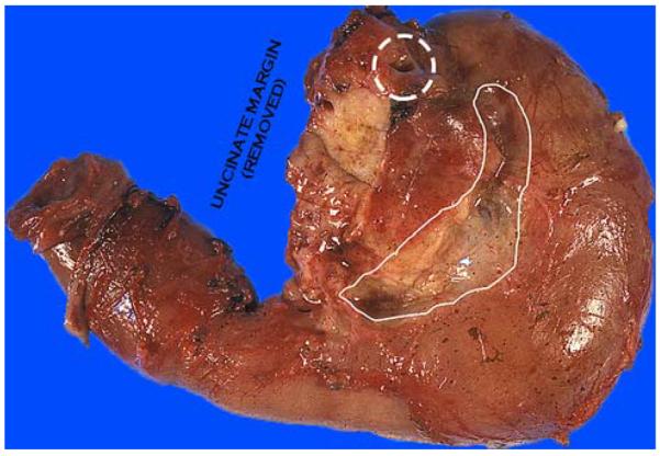
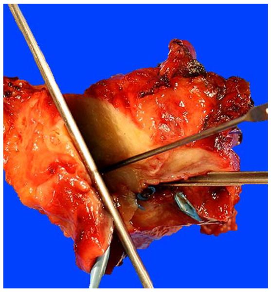
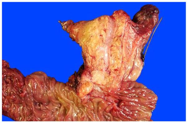
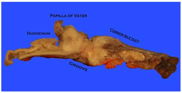
Pancreaticoduodenectomy (PD) specimens present a challenge for surgical pathologists because of the relative rarity of these specimens, combined with the anatomic complexity. Here, we describe our experience on the orientation, dissection, and sampling of PD specimens for a more practical and accurate evaluation of pancreatic, distal common bile duct (CBD), and ampullary tumors. For orientation of PDs, identification of the "trapezoid," created by the vascular bed at the center, the pancreatic neck margin on the left, and the uncinate margin on the right, is of outmost importance in finding all the pertinent margins of the specimen including the CBD, which is located at the upper right edge of this trapezoid. After orientation, all the margins can be sampled. We submit the uncinate margin entirely as a perpendicular inked margin because this adipose tissue-rich area often reveals subtle satellite carcinomas that are grossly invisible, and, with this approach, the number of R1 resections has doubled in our experience. Then, to ensure proper identification of all lymph nodes (LNs), we utilize the orange-peeling approach, in which the soft tissue surrounding the pancreatic head is shaved off in 7 arbitrarily defined regions, which also serve as shaved samples of the so-called "peripancreatic soft tissue" that defines pT3 in the current American Joint Committee on Cancer TNM. With this approach, our LN count increased from 6 to 14 and LN positivity rate from 50% to 73%. In addition, in 90% of pancreatic ductal adenocarcinomas there are grossly undetected microfoci of carcinoma. For determination of the primary site and the extent of the tumor, we believe bisectioning of the pancreatic head, instead of axial (transverse) slicing, is the most revealing approach. In addition, documentation of the findings in the duodenal surface of the ampulla is crucial for ampullary carcinomas and their recent site-specific categorization into 4 categories. Therefore, we probe both the CBD and the pancreatic duct from distal to the ampulla and cut the pancreatic head to the ampulla at a plane that goes through both ducts. Then, we sample the bisected pancreatic head depending on the findings of the case. For example, for proper staging of ampullary carcinomas, it is imperative to take the sections perpendicular to the duodenal serosa at the "groove" area, as ampullary carcinomas often extend to this region. Amputative (axial) sectioning of the ampulla, although good for documentation of the peri-Oddi spread of the intra-ampullary tumors, unfortunately disallows documentation of mucosal spread of the papilla of Vater tumors (those arising from the edge of the ampulla, where the ducts transition to duodenal mucosa and extending) into the neighboring duodenum. Axial sectioning also often fails to document tumor spread to the "groove" area. In conclusion, knowledge of the gross characteristics of the anatomic hallmarks is essential for proper dissection of PD specimens. The approach described above allows practical and accurate documentation and staging of pancreas, distal CBD, and ampullary cancers.


Figures

FIGURE 1
FIGURE 1
Laying the duodenum with the pancreas on top allows readily the identification of the “trapezoid,” located in the postero-median aspect of the pancreatic head. The left vertical edge of the trapezoid is formed by the pancreatic neck margin* (often cauterized, relatively flat and reveals fine granularities) and the right vertical edge by the uncinate margin** (elongated, relatively soft and convex with highly irregular/nodular appearance). A concave-shaped, smooth-surfaced, relatively firm area in between these 2 margins is the vascular bed, where the superior mesenteric vein/portal vein and superior mesenteric artery lie originally.
FIGURE 2
FIGURE 2
The pancreatic duct orifice (green circle), usually located in the upper right quadrant of the pancreatic neck, may be difficult to identify because of its small size. The CBD orifice (red dashed circle) is located at the plateau at the upper right edge of the uncinate margin. It is much larger than the pancreatic duct and should be identifiable in every case. If it has been stitched surgically, the removal of the stitch would make it readily accessible.
FIGURE 3
FIGURE 3
(A) Anterior surface. Anterior surface of a PD typically contains abundant adipose tissue and is convex in appearance. Transition of the pancreatic head to the duodenal wall is also often more irregular, showing a palpable sharp edge due to the overriding pancreatic parenchyma. (B) Posterior surface. In contrast with the anterior surface, the posterior surface is relatively flat, smooth, and glistening. Transition from the pancreatic head to the duodenal wall (pancreaticoduodenal junction) is fairly smooth as well, with the pancreaticoduodenal “groove” only barely identifiable. Note the CBD orifice at the superior edge, at the plateau (arrow).
FIGURE 4
FIGURE 4
PD specimen from the anterior perspective. In this case, the gallbladder and part of the stomach were also removed en bloc. The pancreatic head often has abundant fat in the anterior surface and is localized to the curved part of the “C”-shaped duodenum.
FIGURE 5
FIGURE 5
Our approach to the uncinate margin is to sample it thoroughly as a perpendicular margin, because there is often subtle and nonpalpable invasion into this adipose-rich area. Accordingly, this margin is inked black, cut into a 3 to 5-mm-thick slice (A), bread-loafed, and entirely submitted on edge as a perpendicular margin (B).
FIGURE 6
FIGURE 6
After obtaining the margins, we open the duodenum through the antiampullary (antipancreatic) edge to examine the ampulla. The pancreatic (*) and common bile (**) ducts are probed. There is also a stent (white) in the CBD in this case.
FIGURE 7
FIGURE 7
Although its anatomy varies between individuals, from the duodenal aspect, the major ampulla (dashed circle) is usually readily visible. The accessory ampulla (probe), typically located about 2 cm proximal and slightly anterior to the papilla of Vater, appears as a nodule and can be mistaken for a polyp endoscopically. It is important to examine this area for any mucosal nodularities or irregularities or thickening, as paraduodenal pancreatitis often manifests in this region with variable, often subtle, abnormalities.
FIGURE 8
FIGURE 8
For tumors arising in the ampullary region, careful gross examination of the distribution of the tumor and determination of the preinvasive component (adenomatous lesions that typically manifest as granular, feathery, friable, or smooth-surfaced nodular material; highlighted by gray color in this diagram) are crucial for the proper classification of the ampullary tumors. Ampullary region carcinomas comprise 4 distinct types: Intra-ampullary papillary tubular neoplasm (IAPN)-associated carcinomas are characterized by a significant preinvasive component that grows as an exophytic mass within the ampullary channel (the distal tip of the CBD and the main pancreatic duct). Carcinomas of ampullary ducts are predominantly invasive tumors that circumferentially constrict the distal end of the CBD and pancreatic duct, with minimal changes of the papilla of Vater and ampullary duodenum mucosa. Ampullary duodenal carcinomas usually arise from an adenoma of ampullary duodenum, forming bulky lesions in which the ampullary orifice is often eccentrically located. Tumors that arise from the papilla of Vater itself, as well as those not showing features that characterize the other 3 groups, are classified as ampullary carcinoma, not otherwise specified (NOS).
FIGURE 9
FIGURE 9
The orange-peeling method. The soft tissues covering the pancreas are shaved off after the margins have been obtained and the ampulla examined, before the sectioning of the pancreatic head. This figure illustrates the view after the posterior pancreaticoduodenal LN area (groove between pancreatic head and duodenal wall) has been shaved off. These fragments are searched for LNs and, even if no LN is identified, submitted entirely. These fragments also serve as the documentation of peripancreatic soft tissues, which often yields subtle cancerous foci (isolated solitary ducts). Note the CBD orifice at the superior edge, at the plateau (dashed circle).
FIGURE 10
FIGURE 10
This image illustrates the bivalving of the pancreatic head after both ducts are probed. There is also a stent (blue) in the CBD. With every cut made, the prosector re-checks whether both ducts are still in the same plane. In this case, the knife would have to be re-angled to re-include the CBD.
FIGURE 11
FIGURE 11
Bivalved pancreatic head before fixation. The CBD is significantly larger and light yellow, in the mucosa of which punctate orifices of peribiliary glands are visible on close inspection. The pancreatic duct is usually pink-white with a smooth-glistening appearance and is typically much narrower. Sectioning with this approach allows the evaluation of the compartments of the ampulla (intra-ampullary mucosa, distal segments of the CBD and pancreatic duct, and the duodenal mucosa/wall) as one continuous tube. Thus, distribution of the lesions, not only among these structures but also into their thin walls, can be readily appreciated.
FIGURE 12
FIGURE 12
The ampulla is not covered by the pancreas at the pancreaticoduodenal junction (groove), where ampullary tumors often invade into the periduodenal soft tissues and duodenal serosa without having to go through the pancreas. Therefore, for proper staging of ampullary tumors in this plane, it is important to ink the serosa at the groove area and take perpendicular sections (from tumor to the serosa) to document the tumor spread to this region, which is common in our experience.

Similar articles

Cited by

References

    1. Birkmeyer JD, Siewers AE, Finlayson EV, et al. Hospital volume and surgical mortality in the United States. N Engl J Med. 2002;346:1128–1137. - PubMed
    1. Finks JF, Osborne NH, Birkmeyer JD. Trends in hospital volume and operative mortality for high-risk surgery. N Engl J Med. 2011;364:2128–2137. - PMC - PubMed
    1. Bruzoni M, Johnston E, Sasson AR. Pancreatic incidentalomas: clinical and pathologic spectrum. Am J Surg. 2008;195:329–332. discussion 332. - PubMed
    1. Hopt U, Keck T. Pancreatic incidentalomas. Correct assessment and therapy. Chirurg. 2007;78:713–720. - PubMed
    1. Basturk O, Coban I, Adsay NV. Pancreatic cysts: pathologic classification, differential diagnosis, and clinical implications. Arch Pathol Lab Med. 2009;133:423–438. - PubMed

Publication types

MeSH terms