. 2014 Mar 7;289(10):7247-7256.
doi: 10.1074/jbc.M113.539411. Epub 2014 Jan 22.

Endocytosis and vacuolar degradation of the yeast cell surface glucose sensors Rgt2 and Snf3

Affiliations

Endocytosis and vacuolar degradation of the yeast cell surface glucose sensors Rgt2 and Snf3

Adhiraj Roy et al. J Biol Chem. .

Retraction in

Abstract

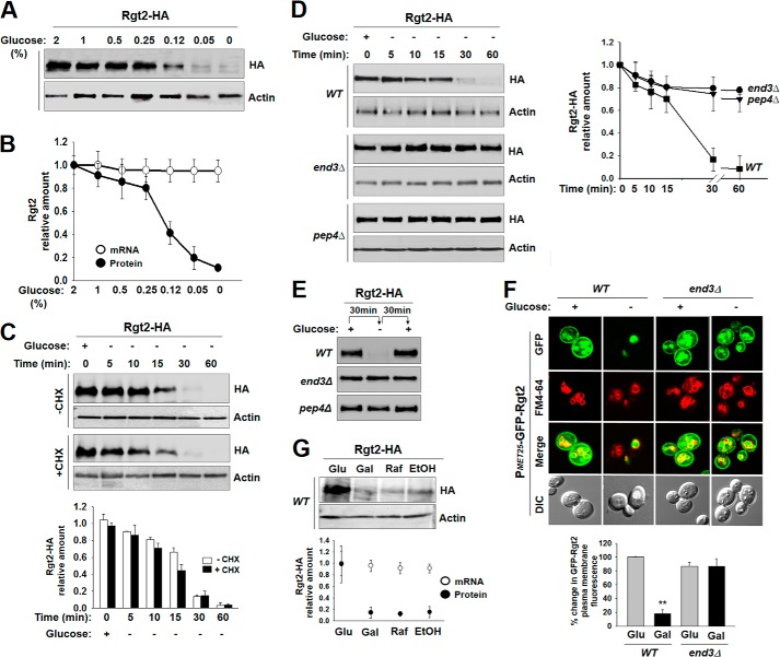
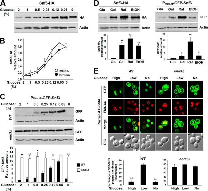
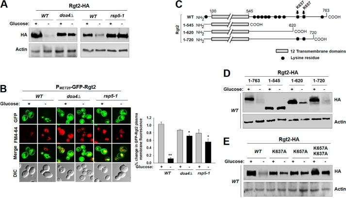
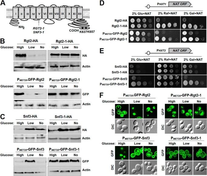
Sensing and signaling the presence of extracellular glucose is crucial for the yeast Saccharomyces cerevisiae because of its fermentative metabolism, characterized by high glucose flux through glycolysis. The yeast senses glucose through the cell surface glucose sensors Rgt2 and Snf3, which serve as glucose receptors that generate the signal for induction of genes involved in glucose uptake and metabolism. Rgt2 and Snf3 detect high and low glucose concentrations, respectively, perhaps because of their different affinities for glucose. Here, we provide evidence that cell surface levels of glucose sensors are regulated by ubiquitination and degradation. The glucose sensors are removed from the plasma membrane through endocytosis and targeted to the vacuole for degradation upon glucose depletion. The turnover of the glucose sensors is inhibited in endocytosis defective mutants, and the sensor proteins with a mutation at their putative ubiquitin-acceptor lysine residues are resistant to degradation. Of note, the low affinity glucose sensor Rgt2 remains stable only in high glucose grown cells, and the high affinity glucose sensor Snf3 is stable only in cells grown in low glucose. In addition, constitutively active, signaling forms of glucose sensors do not undergo endocytosis, whereas signaling defective sensors are constitutively targeted for degradation, suggesting that the stability of the glucose sensors may be associated with their ability to sense glucose. Therefore, our findings demonstrate that the amount of glucose available dictates the cell surface levels of the glucose sensors and that the regulation of glucose sensors by glucose concentration may enable yeast cells to maintain glucose sensing activity at the cell surface over a wide range of glucose concentrations.

Keywords: Cell Biology; Gene Expression; Glucose; Receptors; Yeast.


Figures

FIGURE 1.
FIGURE 1.
Rgt2 undergoes endocytosis and subsequent vacuolar degradation in glucose starved cells. A, Western blot analysis of Rgt2-HA levels at the plasma membrane. Yeast cells (WT) expressing Rgt2-HA were grown in SC-2% glucose medium till mid log phase (A600 nm = 1.2–1.5), and equal amounts of cells were shifted to SC medium containing different concentrations of glucose (0–2%) for 30 min. Membrane fractions were analyzed using anti-HA antibody. B, qRT-PCR analysis of mRNA expression of RGT2 (mRNA) in yeast cells grown as described for Fig. 1A and densitometric quantification of the intensity of each band on the blot in A (Protein). C, yeast cells (WT) expressing Rgt2-HA were grown in SC-2% glucose (+) medium till mid log phase and shifted to 2% galactose (−) medium with or without cycloheximide (CHX, 50 μg/ml) for times as indicated. Membrane fractions were immunoblotted with anti-HA antibody (top panels), and the intensity of each band on the blot was quantified by densitometric scanning (bottom panels). D, yeast cells (WT, end3Δ, and pep4Δ) expressing Rgt2-HA were grown without cycloheximide as described for C. Yeast cells were harvested at different time points as indicated, membrane fractions were immunoblotted with anti-HA antibody (left panel), and the intensity of each band on the blot was quantified by densitometric scanning (bottom panels). E, yeast cells (WT, end3Δ, and pep4Δ) expressing Rgt2-HA were grown in SC-2% glucose medium (+) till mid log phase and shifted to SC-2% galactose medium (−) for 30 min and again shifted to SC-2% glucose medium for 30 min. Membrane fractions were immunoblotted with anti-HA antibody. F, GFP-Rgt2 was expressed from the MET25 promoter in wild type and end3Δ strains. Yeast cells expressing GFP-Rgt2 were grown in SC-2% glucose (+) medium till mid log phase and shifted to 2% galactose (−) medium for 30 min. Confocal microscope images (top panel) and quantification of relative GFP fluorescence in the plasma membrane (bottom panel; **, p < 0.001) were shown. Relative GFP fluorescence intensities were plotted with the fluorescence of WT cells (2% glucose condition) set to 100%. The data represented were averages of at least 50 cell counts with error bars representing S.D. G, yeast cells (WT) expressing Rgt2-HA were grown in SC-2% glucose (Glu) medium till mid log phase and shifted to SC medium containing either 2% galactose (Gal), 2% raffinose (Raf), or 2% ethanol (EtOH) and incubated for 30 min. Membrane fractions were immunoblotted with anti-HA antibody (top panel). qRT-PCR analysis of mRNA expression of RGT2 (mRNA) and densitometric quantification of the intensity of each band on the blot (Protein) (bottom panel). Actin was served as a loading control in A, C, D, E, and G.
FIGURE 2.
FIGURE 2.
Snf3 levels are regulated by both transcriptional and translational mechanisms. A, Western blot analysis of the plasma membrane levels of Snf3-HA. Yeast cells (WT) expressing Snf3-HA were grown as described for Fig. 1A, and membrane fractions were immunoblotted with anti-HA antibody. B, qRT-PCR analysis of mRNA expression of SNF3 (mRNA) in yeast cells grown as described for Fig. 1A and densitometric quantification of the intensity of each band on the blot in A (Protein). C, GFP-Snf3 was expressed from the MET25 promoter in wild type and end3Δ strains. Yeast cells expressing GFP-Snf3 were grown as described for Fig. 1A, and membrane fractions were immunoblotted with anti-HA antibody (top panel). The intensity of each band on the blot was quantified by densitometric scanning (bottom panel; *, p < 0.05; **, p < 0.001). D, Western blot analysis of Snf3-HA and GFP-Snf3 levels at the plasma membrane. Yeast cells (WT) expressing Snf3-HA or GFP-Snf3 were grown as described for Fig. 1G. GFP-Snf3 was expressed from the MET25 promoter. Membrane fractions were immunoblotted with anti-HA antibody (top panels), and the intensity of each band on the blot was quantified by densitometric scanning (bottom panels; *, p < 0.05; **, p < 0.001). E, GFP-Snf3 was expressed from the MET25 promoter in wild type and end3Δ strains in glucose (High), raffinose (Low), or galactose (No) medium. Confocal microscope images (top panel) and quantification of relative GFP fluorescence in the plasma membrane (bottom panels; **, p < 0.001) were shown. Actin was served as a loading control in A, C, and D.
FIGURE 3.
FIGURE 3.
Ubiquitination of the cytoplasmic tail domain of Rgt2 is required for its endocytosis. A, Western blot analysis of Rgt2-HA levels at the plasma membrane. Yeast cells (WT, doa4Δ, and rsp5-1) expressing Rgt2-HA were grown as described for Fig. 1F. Membrane fractions were immunoblotted with anti-HA antibody. B, GFP-Rgt2 was expressed from the MET25 promoter in wild type, doa4Δ, and rsp5-1 strains. Yeast cells expressing GFP-Rgt2 were grown in glucose (+) or galactose (−) medium, as described for Fig. 1F. Confocal microscope images (left panel) and quantification of relative GFP fluorescence in the plasma membrane (right panel; *, p < 0.05; **, p < 0.001) are shown. C, schematic maps of Rgt2 constructs (WT or amino acids 1–545, 1–620, or 1–720) showing lysine residues at N- and C-terminal domains. The Lys637 and Lys657 residues are shown as putative ubiquitin acceptor sites. D and E, yeast cells (WT) expressing the indicated Rgt2-HA constructs were grown as described for Fig. 1F, and membrane fractions were immunoblotted with anti-HA antibody.
FIGURE 4.
FIGURE 4.
Constitutively active Rgt2-1 and Snf3-1 glucose sensors do not undergo endocytosis. A, a schematic diagram of the predicted secondary structure of the Rgt2 glucose sensor showing 12 transmembrane domains, cytoplasmic N- and C-terminal tails, two constitutive mutations (RGT2-1 (R231K) and SNF3-1 (R229K)), and two putative ubiquitin-acceptor lysine residues (Lys637 and Lys657). B and C, yeast cells (WT) expressing the indicated Rgt2 proteins (B) or Snf3 proteins (C) were grown in glucose (High), raffinose (Low), or galactose (No) medium, and membrane fractions were immunoblotted with anti-HA or anti-GFP antibody. Actin served as a loading control. GFP fusions of glucose sensors (GFP-Rgt2, GFP-Rgt2-1, GFP-Snf3, and GFP-Snf3-1) were expressed from the MET25 promoter. D, the PHXT1-NAT reporter strain (JKY88) expressing the indicated Rgt2 proteins was spotted on SC-2% glucose (Glu), SC-2% raffinose (Raf), or SC-2% galactose (Gal) plates supplemented with 100 μg/ml NAT sulfate. The first spot of each row represents a count of 5 × 10⁷ cell/ml, which is diluted 1:10 for each spot thereafter. The glucose plates and the galactose and raffinose plates were incubated for 2 and 3 days, respectively. E, the PHXT2-NAT reporter strain (JKY89) expressing the indicated Snf3 proteins was spotted and photographed as described for D. F, yeast cells (WT) expressing GFP-Rgt2, GFP-Rgt2-1, GFP-Snf3, and GFP-Snf3-1 were grown as described above (B and C) and analyzed by confocal microscopy.
FIGURE 5.
FIGURE 5.
Signaling defective Rgt2 glucose sensor is constitutively endocytosed. A, yeast cells (WT and end3Δ) expressing the indicated Rgt2-HA proteins were grown as described for Fig. 1F, and membrane fractions were immunoblotted with anti-HA antibody (top panels). The intensity of each band on the blot was quantified by densitometric scanning (bottom panel; *, p < 0.05; **, p < 0.001). B, yeast cells (rgt2Δsnf3Δ) expressing the indicated Rgt2-HA proteins were grown as described for Fig. 3C, and the mRNA levels of HXT1 were quantified by qRT-PCR. The values shown are means ± S.D. (*, p < 0.05; **, p < 0.001). C, yeast cells (rgt2Δsnf3Δ) expressing the indicated Rgt2-HA proteins were spotted on 2% glucose plate supplemented with antimycin A (1 μg/ml) (2% Glu + AA) or SC-2% galactose plate (2% Gal) and photographed as described for Fig. 4D. D, yeast cells (rgt2Δsnf3Δ) coexpressing Mth1-Myc and the indicated Rgt2-HA proteins were grown as described for Fig. 1F, and cell lysates were immunoblotted with anti-Myc antibody (top left panels, Mth1-Myc). Actin was served as a loading control (top right panels, actin). Quantification data of Mth1-Myc protein by densitometry are shown (bottom panel; *, p < 0.05; **, p < 0.001).
FIGURE 6.
FIGURE 6.
The turnover of the glucose sensors plays an important role in the adaptation to changing glucose levels. A, a comparison of the amounts of Rgt2-HA and Snf3-HA at the plasma membrane in cells grown in different glucose concentrations is shown (adapted from Figs. 1A and 2A). The first through seventh lanes denote different concentrations of glucose (%, w/v): 2, 1, 0.5, 0.25, 0.125, 0.05, and 0, respectively. B, stability of the glucose sensors is associated with their ability to sense glucose. The low affinity glucose sensor Rgt2 is endocytosed and targeted to the vacuole for degradation in glucose-starved cells but is stable in cells grown in high glucose medium. Expression of the high affinity glucose sensor Snf3 is regulated at both transcriptional and posttranslational levels: Snf3 is internalized and degraded not only in high glucose-grown cells but also in glucose-depleted cells; its expression is repressed by high glucose concentrations but derepressed in the absence of glucose. Consequently, Snf3 is stable in glucose-starved cells. Regulation of the turnover of the glucose sensors enables yeast cells to produce the glucose transporters most appropriate for the amount of glucose available in the environment.

Similar articles

Cited by

References

    1. Rolland F., Wanke V., Cauwenberg L., Ma P., Boles E., Vanoni M., de Winde J. H., Thevelein J. M., Winderickx J. (2001) The role of hexose transport and phosphorylation in cAMP signalling in the yeast Saccharomyces cerevisiae. FEMS Yeast Res. 1, 33–45 - PubMed
    1. Towle H. C. (2005) Glucose as a regulator of eukaryotic gene transcription. Trends Endocrinol. Metab. 16, 489–494 - PubMed
    1. Ozcan S., Johnston M. (1999) Function and regulation of yeast hexose transporters. Microbiol. Mol. Biol. Rev. 63, 554–569 - PMC - PubMed
    1. Holsbeeks I., Lagatie O., Van Nuland A., Van de Velde S., Thevelein J. M. (2004) The eukaryotic plasma membrane as a nutrient-sensing device. Trends Biochem. Sci. 29, 556–564 - PubMed
    1. Johnston M., Kim J. H. (2005) Glucose as a hormone. Receptor-mediated glucose sensing in the yeast Saccharomyces cerevisiae. Biochem. Soc. Trans. 33, 247–252 - PubMed

Publication types

MeSH terms

Substances