. 2014 Mar 7;289(10):6991-7002.
doi: 10.1074/jbc.M113.511444. Epub 2014 Jan 22.

Histone deacetylase inhibitors modulate the transcriptional regulation of guanylyl cyclase/natriuretic peptide receptor-a gene: interactive roles of modified histones, histone acetyltransferase, p300, AND Sp1

Affiliations

Histone deacetylase inhibitors modulate the transcriptional regulation of guanylyl cyclase/natriuretic peptide receptor-a gene: interactive roles of modified histones, histone acetyltransferase, p300, AND Sp1

Prerna Kumar et al. J Biol Chem. .

Abstract

Atrial natriuretic peptide (ANP) binds guanylyl cyclase-A/natriuretic peptide receptor-A (GC-A/NPRA) and produces the intracellular second messenger, cGMP, which regulates cardiovascular homeostasis. We sought to determine the function of histone deacetylases (HDACs) in regulating Npr1 (coding for GC-A/NPRA) gene transcription, using primary mouse mesangial cells treated with class-specific HDAC inhibitors (HDACi). Trichostatin A, a pan inhibitor, and mocetinostat (MGCD0103), a class I HDAC inhibitor, significantly enhanced Npr1 promoter activity (by 8- and 10-fold, respectively), mRNA levels (4- and 5.3-fold, respectively), and NPRA protein (2.7- and 3.5-fold, respectively). However, MC1568 (class II HDAC inhibitor) had no discernible effect. Overexpression of HDAC1 and HDAC2 significantly attenuated Npr1 promoter activity, whereas HDAC3 and HDAC8 had no effect. HDACi-treated cultured cells in vitro and intact animals in vivo showed significantly reduced binding of HDAC1 and -2 and increased accumulation of acetylated H3-K9/14 and H4-K12 at the Npr1 promoter. Deletional analyses of the Npr1 promoter along with ectopic overexpression and inhibition of Sp1 confirmed that HDACi-induced Npr1 gene transcription is accomplished by Sp1 activation. Furthermore, HDACi attenuated the interaction of Sp1 with HDAC1/2 and promoted Sp1 association with p300 and p300/cAMP-binding protein-associated factor; it also promoted the recruitment of p300 and p300/cAMP-binding protein-associated factor to the Npr1 promoter. Our results demonstrate that trichostatin A and MGCD0103 enhanced Npr1 gene expression through inhibition of HDAC1/2 and increased both acetylation of histones (H3-K9/14, H4-K12) and Sp1 by p300, and their recruitment to Npr1 promoter. Our findings define a novel epigenetic regulatory mechanism that governs Npr1 gene transcription.

Keywords: Chromatin Histone Modification; Gene Transcription; Histone Deacetylase Inhibitors; Natriuretic Peptides; Sp1; p300.


Figures
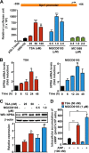
FIGURE 1.
FIGURE 1.
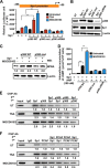
Effect of TSA and MGCD0103 on Npr1 gene transcription and expression. A, luciferase activity of the Npr1 proximal promoter construct −356/+55 in MMCs treated with increasing concentrations of HDACi (TSA, MGCD0103, and MC1568). B, effect of TSA (50 nm) and MGCD0103 (1 μm) on Npr1 mRNA levels in a time-dependent manner as determined by real-time RT-PCR. C, Western blot (WB) analysis of NPRA protein expression in cells treated with HDACi and β-actin expression is shown as the loading control. D, intracellular accumulation of cGMP in MMCs treated with HDACi (TSA, 50 nm; MGCD0103, 1 μm) and ANP. Bar represents the mean ± S.E. of four independent experiments in triplicates. WB, Western blot; *, p < 0.05; **, p < 0.01; ***, p < 0.001.
FIGURE 2.
FIGURE 2.
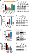
Effect of the overexpression and knockdown of HDAC1 and HDAC2 in transcriptional regulation and expression of the Npr1 gene. A, luciferase activity of Npr1 proximal promoter (−356/+55) in MMCs cotransfected with class I HDACs (HDAC1, -2, -3, and -8) or an empty vector. B, Western blot (WB) analysis of HDAC1, -2, and -3 in transfected cells. β-Actin was used as loading control. C, luciferase activity of the Npr1 promoter cotransfected with HDAC1, HDAC2, and control siRNA (0.1 μm). D, Western blot analysis of siRNA-mediated knockdown of endogenous HDAC1 and HDAC2 proteins in transfected cells with β-actin as loading control. E, luciferase activity of the Npr1 promoter construct −356/+55 cotransfected with catalytically inactive HDAC1 (250 ng) and HDAC2 (250 ng) mutant plasmid. F, Western blot analysis of mutant HDAC1 and -2 proteins in transfected cells with β-actin as loading control. G, Npr1 mRNA levels in cells transfected with HDAC1 and HDAC2 expression plasmid as analyzed by real-time RT-PCR with β-actin as an internal control. H, Western blot analysis of NPRA protein expression in cells transfected with HDAC1 and HDAC2 expression plasmid and β-actin expression is shown as loading control. Data represent mean ± S.E. of four independent experiments. Ctrl, control; UT, untransfected; *, p < 0.05; **, p < 0.01; ***, p < 0.001.
FIGURE 3.
FIGURE 3.
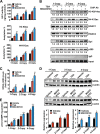
Modulation of total HDAC activity and HDAC1 and HDAC2 protein expression by TSA, MGCD0103, and MC1568. A, total HDAC activity was measured in nuclear extracts of HDACi-treated and untreated MMCs. B, semiquantitative measurement of HDAC1 and HDAC2 protein levels in HDACi-treated cells by colorimetric method. C, Western blot analysis of HDAC1, -2, and -3 protein expressions in HDACi-treated cells with β-actin expression as loading control. D, densitometric analysis for HDAC1, -2, and -3. Bar represents mean ± S.E. of four independent experiments. HDACi (TSA, 50 nm; MGCD0103, 1 μm; MC1568, 1 μm); UT, untreated; WB, Western blot; *, p < 0.05; **, p < 0.01; ***, p < 0.001.
FIGURE 4.
FIGURE 4.
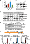
Effect of TSA and MGCD0103 on HAT activity, histone acetylation, and association with the Npr1 promoter. A, quantification of total HAT activity in HDACi-treated MMCs by the colorimetric method. B, Western blot analysis of p300 and PCAF protein expression in HDACi-treated cells. β-Actin was used as loading control. Concentration (C)- and time-dependent (D) effect of HDACi on histones H3-K9/14ac, H4-K12ac, and H3-K9me3 protein levels as analyzed by Western blot. Histone H3 was used as loading control. E, schematic map of the Npr1 promoter showing the −1155/−914 bp and −120/+73 bp regions. Quantitative ChIP assay demonstrating in vivo accumulation of acetylated histones to the Npr1 promoter region. Bar represents mean ± S.E. of three independent experiments. HDACi (TSA, 50 nm; MGCD0103, 1 μm); UT, untreated; Ab, antibody; WB, Western blot; *, p < 0.05; **, p < 0.01; ***, p < 0.001.
FIGURE 5.
FIGURE 5.
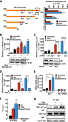
Role of Sp1 in mediating TSA and MGCD0103 effects on Npr1 gene transcription. A, schematic map of the Npr1 promoter deletion construct and their luciferase activity in transfected MMCs treated with HDACi. B, luciferase activity of the Npr1 promoter construct −356/+55 cotransfected with Sp1 plasmid and treated with HDACi. Blots show Western blot analysis of Sp1 protein expression in HDACi-treated cells with β-actin as loading control. C, luciferase activity of the Npr1 promoter cotransfected with Sp1 siRNA and treated with HDACi. Lower panel, Western blot analysis of Sp1 protein expression in Sp1 siRNA-transfected cells and β-actin as loading control. D, luciferase activity of the Npr1 promoter in cells pretreated with mithramycin A and induced with HDACi. E, effect of HDACi on Npr1 mRNA levels in cells transfected with Sp1 expression plasmid or Sp1 siRNA as determined by real-time RT-PCR with β-actin as the internal control. F, quantitative ChIP assay demonstrating recruitment of Sp1 protein on the Npr1 promoter (−120 to +73) in HDACi-stimulated cells as determined by real-time PCR. G, Western blot analysis of acetylated and total Sp1 in the immunoprecipitate from HDACi-treated cells. Input shows Sp1 in lysates as detected by Western blot. Bars represent mean ± S.E. of three independent experiments. HDACi (TSA, 50 nm; MGCD0103, 1 μm); Ctrl, control; UT, untreated; mith A, mithramycin A; WB, Western blot; IP, immunoprecipitation; *, p < 0.05; **, p < 0.01; ***, p < 0.001.
FIGURE 6.
FIGURE 6.
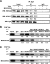
Effect of HDACi on interaction of HDAC1 and -2 with Sp1 on the Npr1 promoter. A, Western blot analysis of HDAC1, HDAC2, or Sp1 complexes isolated by immunoprecipitation with anti-Sp1 antibody from HDACi-treated cells. Sequential ChIP analysis demonstrating in vivo recruitment of HDAC1 (B) and HDAC2 (C) to the Npr1 promoter by Sp1 in MGCD0103-treated and untreated cells. The intensity of DNA bands was quantified by Alpha Innotech analysis software. Data shown represent mean ± S.E. of three independent experiments. MGCD0103 (1 μm); UT, untreated; WB, Western blot; IP, immunoprecipitation.
FIGURE 7.
FIGURE 7.
Interaction of p300 and PCAF with Sp1 in the HDACi-mediated effect on Npr1 gene transcription and expression. A, luciferase activity of Npr1 promoter construct −356/+55 cotransfected with Sp1 and wild-type p300, mutant p300-HAT, or p300 siRNA expression plasmid and treated with HDACi. B, Western blot analysis of Sp1, wild-type p300, mutant p300-HAT, and siRNA-mediated knockdown of endogenous p300 in transfected cells. β-Actin was used as loading control. C, Western blot analysis of NPRA protein expression in cells overexpressing Sp1 and wild-type p300 or mutant p300-HAT in the presence of MGCD0103 and β-actin expression is shown as loading control. D, intracellular accumulation of cGMP in cells overexpressing wild-type or mutant p300 and Sp1 protein treated with MGCD0103 and induced with ANP. Sequential ChIP analysis demonstrating in vivo recruitment of p300 (E) and PCAF (F) to the Npr1 promoter by Sp1 in HDACi-treated and untreated cells. The intensity of DNA bands was quantified by Alpha Innotech analysis software. Representative gels from three independent experiments are shown. Data shown represent mean ± S.E. of three independent experiments. HDACi (TSA, 50 nm; MGCD0103, 1 μm); Ctrl, control; UT, untreated; WB, Western blot; IP, immunoprecipitation; **, p < 0.01.
FIGURE 8.
FIGURE 8.
HDACi-mediated in vivo regulation of renal NPRA expression and signaling involving HDACs, acetylated histones, Sp1, and p300 in Npr1 gene-targeted mice. A, effect of HDACi on renal HAT activity and global acetylation levels of H3-K9ac and H4-K12ac. B, HDACi-mediated recruitment of acetylated histones, HDAC1 and -2, Sp1, and p300 at the Npr1 promoter (−120 to +73) in treated mice kidneys. Pooled samples from 6 mice in each group were used for ChIP and purified DNA was amplified by conventional PCR. C, Npr1 mRNA levels in TSA- and MGCD0103-treated mice kidneys as analyzed by real-time RT-PCR with β-actin as an internal control. D, Western blot and densitometry analyses of NPRA protein expression in TSA- and MGCD0103-treated Npr1 1-copy, 2-copy, and 4-copy mice kidneys. β-Actin was used as loading control. E, cGMP levels in kidney tissues of HDACi-treated mice as quantitated by ELISA. Data shown represent mean ± S.E. of three independent experiments. *, p < 0.05; **, p < 0.01; ***, p < 0.001 (vehicle-treated versus drug-treated same group). Ab, antibody. In each group, six mice (n = 6) were used.
FIGURE 9.
FIGURE 9.
Schematic model showing TSA- and MGCD0103-mediated regulation of Npr1 gene transcription and expression via interactions among regulatory elements and chromatin remodeling. The schematic model depicts that under untreated conditions class I HDACs (HDAC1 and -2) interact with Sp1 and form a corepressor complex and also maintain H3 and H4 in a hypoacetylated state. Under these conditions, transcription factors, including Sp1 has low affinity for promoter. Treatment with HDACi (TSA and MGCD0103) attenuates HDAC activity and induces HAT activity. This allows HAT proteins, p300 and PCAF, whose expression was induced by TSA and MGCD0103, to hyperacetylate H3 at K9/14, H4 at K12, and Sp1, which in association with p300 and PCAF occupies the Npr1 promoter within the newly formed permissive chromatin condition and leads to increased Npr1 gene transcription.

Similar articles

Cited by

References

    1. Drewett J. G., Garbers D. L. (1994) The family of guanylyl cyclase receptors and their ligands. Endocr. Rev. 15, 135–162 - PubMed
    1. de Bold A. J. (1985) Atrial natriuretic factor. A hormone produced by the heart. Science 230, 767–770 - PubMed
    1. Pandey K. N., Singh S. (1990) Molecular cloning and expression of murine guanylate cyclase/atrial natriuretic factor receptor cDNA. J. Biol. Chem. 265, 12342–12348 - PubMed
    1. Levin E. R., Gardner D. G., Samson W. K. (1998) Natriuretic peptides. N. Engl. J. Med. 339, 321–328 - PubMed
    1. Pandey K. N. (2005) Biology of natriuretic peptides and their receptors. Peptides 26, 901–932 - PubMed

Publication types

MeSH terms

LinkOut - more resources