. 2014 Mar 14;289(11):7335-48.
doi: 10.1074/jbc.M113.545046. Epub 2014 Jan 22.

Characterization of all family-9 glycoside hydrolases synthesized by the cellulosome-producing bacterium Clostridium cellulolyticum

Affiliations

Characterization of all family-9 glycoside hydrolases synthesized by the cellulosome-producing bacterium Clostridium cellulolyticum

Julie Ravachol et al. J Biol Chem. .

Abstract

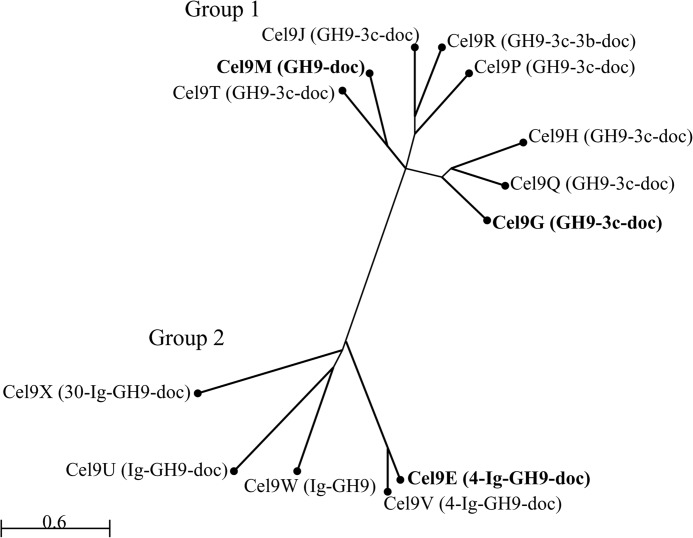
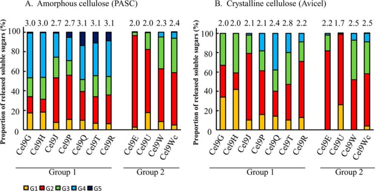
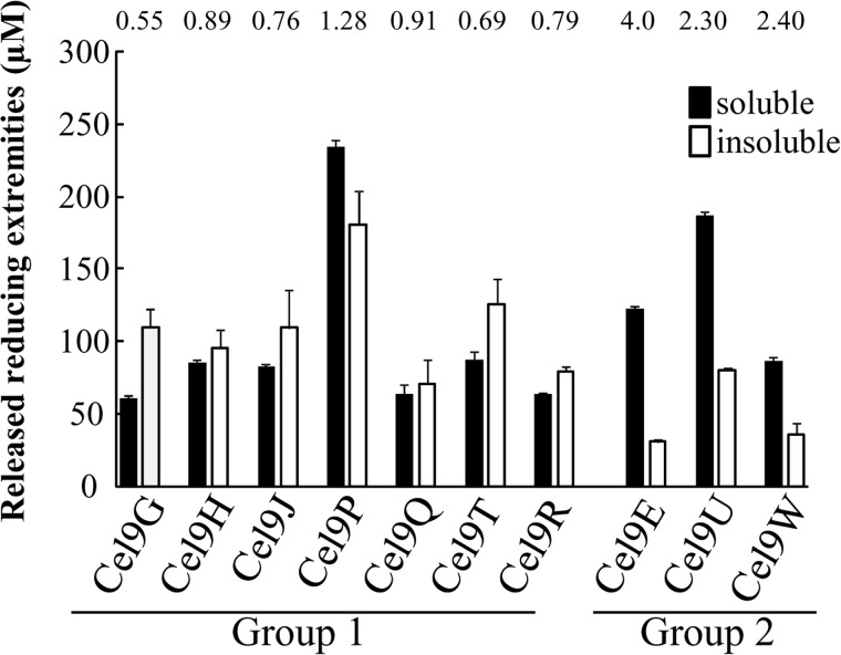
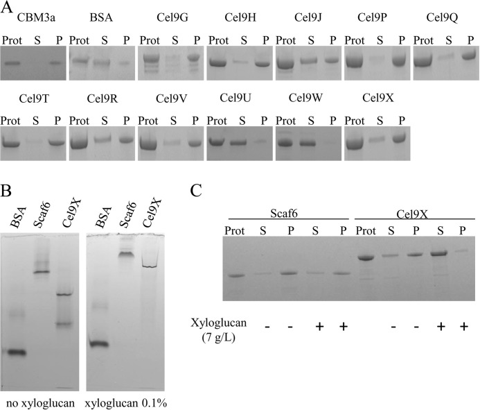
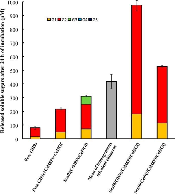
The genome of Clostridium cellulolyticum encodes 13 GH9 enzymes that display seven distinct domain organizations. All but one contain a dockerin module and were formerly detected in the cellulosomes, but only three of them were previously studied (Cel9E, Cel9G, and Cel9M). In this study, the 10 uncharacterized GH9 enzymes were overproduced in Escherichia coli and purified, and their activity pattern was investigated in the free state or in cellulosome chimeras with key cellulosomal cellulases. The newly purified GH9 enzymes, including those that share similar organization, all exhibited distinct activity patterns, various binding capacities on cellulosic substrates, and different synergies with pivotal cellulases in mini-cellulosomes. Furthermore, one enzyme (Cel9X) was characterized as the first genuine endoxyloglucanase belonging to this family, with no activity on soluble and insoluble celluloses. Another GH9 enzyme (Cel9V), whose sequence is 78% identical to the cellulosomal cellulase Cel9E, was found inactive in the free and complexed states on all tested substrates. The sole noncellulosomal GH9 (Cel9W) is a cellulase displaying a broad substrate specificity, whose engineered form bearing a dockerin can act synergistically in minicomplexes. Finally, incorporation of all GH9 cellulases in trivalent cellulosome chimera containing Cel48F and Cel9G generated a mixture of heterogeneous mini-cellulosomes that exhibit more activity on crystalline cellulose than the best homogeneous tri-functional complex. Altogether, our data emphasize the importance of GH9 diversity in bacterial cellulosomes, confirm that Cel9G is the most synergistic GH9 with the major endoprocessive cellulase Cel48F, but also identify Cel9U as an important cellulosomal component during cellulose depolymerization.

Keywords: Carbohydrate; Cellulase; Cellulose; Cellulosome; Clostridium cellulolyticum; Enzyme Kinetics; Enzymes; GH9; Protein Complexes; Synergy.


Figures

FIGURE 1.
FIGURE 1.
Schematic representation of the recombinant proteins used in this study. The GH and CBM families are indicated. Underlined names correspond to the enzymes produced, purified, and characterized in this study. Cel48Ft designates Cel48F from C. cellulolyticum bearing a C. thermocellum dockerin. Cel9Gf designates Cel9G from C. cellulolyticum appended with a R. flavefaciens dockerin. Cel9Wc refers to an engineered form of Cel9W appended with a C. cellulolyticum dockerin.
FIGURE 2.
FIGURE 2.
Phylogenic analysis of the catalytic domains of the GH9 enzymes from C. cellulolyticum. The phylogenetic tree was generated using neighbor joining analyses based on COBALT alignment of the GH9 catalytic domain sequences. Proteins in bold correspond to enzymes that were formerly characterized. The modular organization of each enzyme is indicated as follows: GH9, glycoside hydrolase family 9 catalytic domain; doc, dockerin; Ig, immunoglobulin like module; 3c, 3b, 4, and 30, family 3c, 3b, 4, and 30 carbohydrate binding module, respectively. Scale bar corresponds to 0.6% amino acid substitution.
FIGURE 3.
FIGURE 3.
Analysis of the cellodextrins released by the various GH9 cellulases on insoluble celluloses. Soluble sugars generated by 0.1 μm of enzyme after 30 min of incubation on amorphous cellulose (A) or 24 h on crystalline cellulose (B) were identified and quantified by HPAEC-PAD. Numbers on top of the bars designate the average degree of polymerization of the released cellodextrins.
FIGURE 4.
FIGURE 4.
Mode of action of the various GH9 cellulases on amorphous cellulose. The amount of soluble and insoluble reducing extremities generated by each enzyme on 3.5 g/liter amorphous cellulose is reported. Numbers on top of the bars indicate the soluble-to-insoluble ratios. The data show the mean and standard deviation of three independent experiments.
FIGURE 5.
FIGURE 5.
Xyloglucan degradation patterns by Cel9U and Cel9X. The samples were analyzed by HPAEC-PAD. Black line, xyloglucan at 3.5 g/liter incubated for 2 h at 37 °C; red line, xyloglucan (3.5 g/liter) incubated for 2 h at 37 °C with 10 nm of Cel9U; blue line, xyloglucan (3.5 g/liter) incubated for 30 min at 37 °C with 10 nm of Cel9X. XGOGlc4, XGOGlc8, and XGOGlc24) refer to xyloglucan oligosaccharides displaying 4, 8, and 24 glucosyl residues backbone, respectively. G designates unbranched β(1→4)-linked backbone glucosyl residue. The X unit represents a (Xylα(1→6))Glc β(1→4) moiety. The L unit refers to ([Galβ(1→2))Xylα(1→6)]Glc β(1→4) moiety (27).
FIGURE 6.
FIGURE 6.
Far-UV CD spectra of Cel9E and Cel9V. Spectra were recorded at 25 °C in 1-mm path length quartz cell. Protein concentration was 1 μm. Red line, Cel9E; black line, Cel9V purified by anion exchange chromatography (final purification step); blue line, Cel9V purified using gel filtration chromatography (final purification step).
FIGURE 7.
FIGURE 7.
Binding of GH9 enzymes to cellulosic substrates. The binding to amorphous cellulose (A), xyloglucan (B), and both (C) were investigated. A, proteins (1.5 μm) were mixed with amorphous cellulose (7 g/liter) and incubated for 1 h at 4 °C. The suspension was centrifuged; the pellet (P, bound proteins) was washed twice, and the supernatant fluids (S, unbound protein) were collected, mixed with sample buffer, and subjected to SDS-PAGE, together with an aliquot of the protein solution prior incubation with the substrate (Prot). CBM3a designates the CBM3a of the scaffoldin CipC from C. cellulolyticum produced in E. coli. B, nondenaturing electrophoresis of BSA, Scaf6, and Cel9X on polyacrylamide gel containing no or 0.1% (w/v) xyloglucan. C, binding assays of Scaf6 and Cel9X onto amorphous cellulose in the presence or absence of xyloglucan (7 g/liter). Prot, S, and P same as in Fig. 6A.
FIGURE 8.
FIGURE 8.
Degradation of crystalline cellulose by a mixture of 11 Scaf6-based trivalent chimeras containing Cel48Ft, Cel9Gf, and one GH9 cellulase bearing a C. cellulolyticum dockerin. The amount of released soluble cellodextrins and their proportion after 24 h of incubation at 37 °C with 3.5 g/liter Avicel are shown. Free GH9s refers to a mixture of free Cel9E + Cel9G + Cel9H + Cel9J + Cel9M + Cel9P + Cel9Q + Cel9R + Cel9T + Cel9U + Cel9Wc, each enzyme being at a final concentration of 0.0091 μm. Free GH9s + Cel48Ft + Cel9Gf refers to free GH9s (each enzyme at 0.0091 μm) supplemented with free Cel48Ft and Cel9Gf at 0.1 μm. Scaf6(Cel48Ft/Cel9Gf) refers to the Scaf6-based divalent complex at 0.1 μm with no enzyme bound onto the C. cellulolyticum cohesin of the scaffoldin. Mean of homogeneous trivalent chimeras corresponds to the average activity on Avicel of all homogenous trivalent chimeras at 0.1 μm containing Cel48Ft, Cel9Gf, and one GH9 cellulase, which was calculated from the data reported in Table 5. Scaf6(GH9s/Cel48Ft/Cel9Gf) designates a mixture of the Scaf6-based complexes that systematically contain Cel48Ft and Cel9Gf and either Cel9E, Cel9G, Cel9H, Cel9J, Cel9M, Cel9P, Cel9Q, Cel9R, Cel9T, Cel9U, or Cel9Wc bound onto the C. cellulolyticum cohesin of the scaffoldin. Each distinct trivalent complex in the mixture was at a concentration of 0.0091 μm. Scaf6(Cel9U/Cel48Ft/Cel9Gf) refers to the best homogeneous trivalent chimera (see Table 5), which was at a concentration of 0.1 μm. The data show the mean of at least three independent experiments, and bars indicate the standard deviation.

Similar articles

Cited by

References

    1. Cantarel B. L., Coutinho P. M., Rancurel C., Bernard T., Lombard V., Henrissat B. (2009) The carbohydrate-active EnZymes database (CAZy): an expert resource for glycogenomics. Nucleic Acids Res. 37, D233–D238 - PMC - PubMed
    1. Aspeborg H., Coutinho P. M., Wang Y., Brumer H., 3rd, Henrissat B. (2012) Evolution, substrate specificity and subfamily classification of glycoside hydrolase family-5 (GH5). BMC Evol. Biol. 12, 186. - PMC - PubMed
    1. Henrissat B., Claeyssens M., Tomme P., Lemesle L., Mornon J. P. (1989) Cellulase families revealed by hydrophobic cluster analysis. Gene 81, 83–95 - PubMed
    1. Honda Y., Shimaya N., Ishisaki K., Ebihara M., Taniguchi H. (2011) Elucidation of exo-β-d-glucosaminidase activity of a family 9 glycoside hydrolase (PBPRA0520) from Photobacterium profundum SS9. Glycobiology 21, 503–511 - PubMed
    1. Arai T., Araki R., Tanaka A., Karita S., Kimura T., Sakka K., Ohmiya K. (2003) Characterization of a cellulase containing a family 30 carbohydrate-binding module (CBM) derived from Clostridium thermocellum CelJ: importance of the CBM to cellulose hydrolysis. J. Bacteriol. 185, 504–512 - PMC - PubMed

Publication types

LinkOut - more resources