. 2014 Mar 14;289(11):7293-306.
doi: 10.1074/jbc.M113.520403. Epub 2014 Jan 22.

A redox-resistant sirtuin-1 mutant protects against hepatic metabolic and oxidant stress

Affiliations

A redox-resistant sirtuin-1 mutant protects against hepatic metabolic and oxidant stress

Di Shao et al. J Biol Chem. .

Abstract

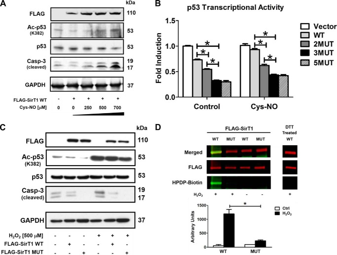
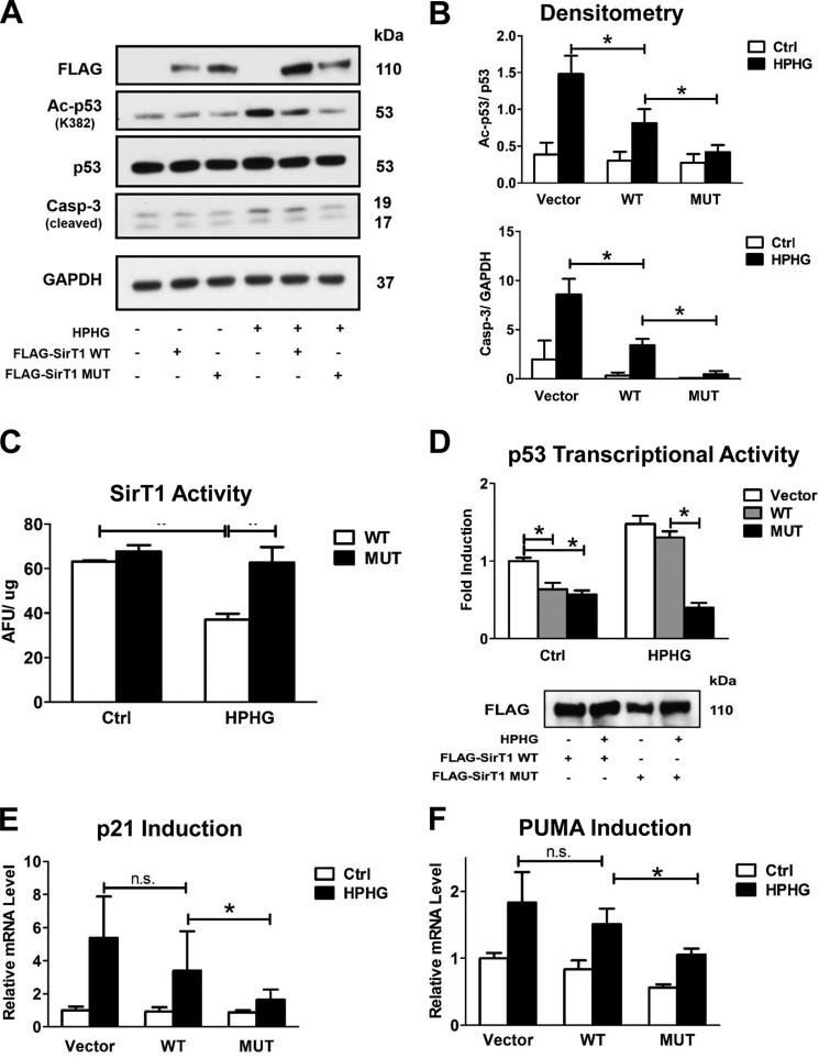
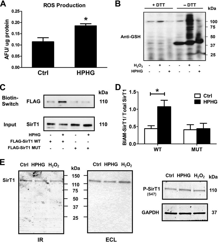
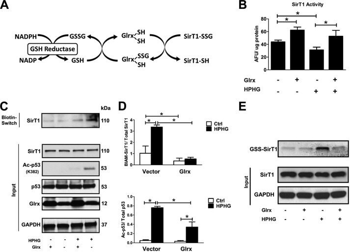
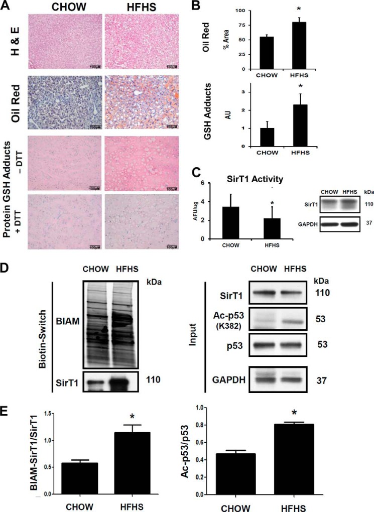
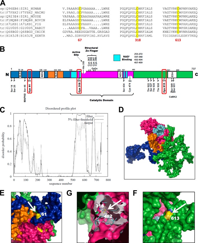
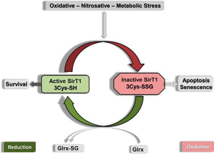
Sirtuin-1 (SirT1), a member of the NAD(+)-dependent class III histone deacetylase family, is inactivated in vitro by oxidation of critical cysteine thiols. In a model of metabolic syndrome, SirT1 activation attenuated apoptosis of hepatocytes and improved liver function including lipid metabolism. We show in SirT1-overexpressing HepG2 cells that oxidants (nitrosocysteine and hydrogen peroxide) or metabolic stress (high palmitate and high glucose) inactivated SirT1 by reversible oxidative post-translational modifications (OPTMs) on three cysteines. Mutating these oxidation-sensitive cysteines to serine preserved SirT1 activity and abolished reversible OPTMs. Overexpressed mutant SirT1 maintained deacetylase activity and attenuated proapoptotic signaling, whereas overexpressed wild type SirT1 was less protective in metabolically or oxidant-stressed cells. To prove that OPTMs of SirT1 are glutathione (GSH) adducts, glutaredoxin-1 was overexpressed to remove this modification. Glutaredoxin-1 overexpression maintained endogenous SirT1 activity and prevented proapoptotic signaling in metabolically stressed HepG2 cells. The in vivo significance of oxidative inactivation of SirT1 was investigated in livers of high fat diet-fed C57/B6J mice. SirT1 deacetylase activity was decreased in the absence of changes in SirT1 expression and associated with a marked increase in OPTMs. These results indicate that glutathione adducts on specific SirT1 thiols may be responsible for dysfunctional SirT1 associated with liver disease in metabolic syndrome.

Keywords: Glutathiolation; Glutathionylation; High Fat High Sucrose Diet; Metabolic Diseases; Oxidative Stress; Polyphenols; Reactive Oxygen and Nitrogen Species; Resveratrol.


Figures

FIGURE 1.
FIGURE 1.
SirT1 WT inactivation by oxidative and nitrosative stress is abrogated by a triple cysteine mutant in HepG2 cells. HepG2 cells were transiently transfected for 48 h with either FLAG-SirT1 WT, FLAG-SirT1 MUT (mutated residues C61S, C318S, and C613S), or empty vector (pcDNA3.1) as a control. Equal SirT1 overexpression was confirmed by Western blot analysis with an antibody against the FLAG tag. Representative Western blots of three independent experiments are shown. A, cells were treated with Cys-NO (bolus addition) for 8 h at the indicated concentrations. Overexpression of SirT1 suppressed caspase 3 (Casp-3) cleavage and p53 acetylation (Ac-p53) at the SirT1-specific substrate site Lys382. Increased concentrations of Cys-NO inhibited the SirT1-dependent effect, increasing p53 acetylation and proapoptotic cleaved caspase 3. B, p53 transcriptional activity was measured by transfecting a PUMA-specific luciferase reporter into cells. Cys-NO (bolus addition of 500 μm) treatment inhibited the SirT1-dependent (WT) suppression of PUMA promoter activity, but the double, triple, and quintuple mutants (2MUT, 3MUT, and 5MUT) maintained PUMA suppression when treated with Cys-NO. The triple mutant (3MUT) exhibited the greatest effect and was used for the remainder of the experiments (referred to as MUT SirT1). ANOVA and Bonferroni's post-test were used (n = 3). Error bars, S.D. *, p < 0.001, WT SirT1- versus SirT1 mutant-transfected HepG2 cells either under control conditions or treated with Cys-NO. C, cells were treated with H2O2 (500 μm bolus addition) for 8 h. Overexpression of SirT1 WT reduced cleaved caspase 3 in H2O2-treated cells, whereas MUT almost restored control levels. D, reversible oxidative modifications of cysteines were detected in SirT1 WT and MUT with a biotin switch assay and IR-based (LI-COR Biosciences) Western analysis. Transfected cells were treated for 8 h with hydrogen peroxide (bolus addition) (n = 3). Cysteines of WT SirT1 were reversibly modified by H2O2 (500 μm) treatment as demonstrated by HPDP-biotin labeling of the reduced cysteine modification (green, biotin-labeled protein; red, FLAG antibody for detection of SirT1). DTT treatment preceding the labeling procedure (right panel) shows no staining, confirming the specificity of the method for reversible oxidation. For details please refer to the “Experimental Procedures.” ANOVA and Bonferroni's post-test were used (n = 3). Error bars, S.D. *, p < 0.001, WT versus MUT SirT1-transfected HepG2 cells treated with H2O2.
FIGURE 2.
FIGURE 2.
SirT1 MUT maintained activity and prevented proapoptotic signaling in HPHG-treated HepG2 cells. HepG2 cells were transiently transfected for 48 h with either FLAG-SirT1 WT, FLAG-SirT1 MUT (mutated residues C61S, C318S, and C613S), or empty vector (pcDNA3.1) as a control. SirT1 overexpression was confirmed by Western blot analysis with an antibody against the FLAG tag. A, overexpression of SirT1 WT suppressed p53 acetylation (Ac-p53) at the SirT1-specific substrate site Lys382 and caspase 3 (Casp-3) cleavage in normal medium. In contrast, treating cells with HPHG for 16 h inhibited the SirT1-dependent effect, increasing p53 acetylation and proapoptotic cleaved caspase 3. The SirT1 MUT, however, maintained control levels. B, densitometric analysis of the Western blot results normalized to GAPDH for cleaved caspase 3 or total p53 for acetylated p53. ANOVA and Bonferroni's post-test were used (n = 3). Error bars, S.D. *, p < 0.001, WT SirT1 versus empty vector control; *, p < 0.05, WT SirT1- versus MUT SirT1-transfected HepG2 cells treated with HPHG. C, overexpressed FLAG-SirT1 WT or MUT was either isolated from transfected control or cells treated with HPHG. Activity of the isolated enzyme was measured by the Fluor-de-Lys assay. HPHG treatment decreased SirT1 WT, but MUT activity was fully maintained. ANOVA and Bonferroni's post-test were used (n = 3). Error bars, S.D. *, p < 0.001, WT SirT1- versus SirT1 MUT-transfected HepG2 cells treated with HPHG. D, p53 transcriptional activity was measured by transfecting a PUMA-specific luciferase reporter together with FLAG-SirT1 WT, FLAG-SirT1 MUT, or empty vector (pcDNA3.1) into cells for 48 h. HPHG treatment for 16 h inhibited the SirT1-dependent suppression of PUMA promoter activity, but the MUT maintained PUMA suppression when cells were exposed to HPHG. ANOVA and Bonferroni's post-test were used (n = 3). Error bars, S.D. *, p < 0.001, empty vector versus WT SirT1 and empty vector- versus SirT1 MUT-transfected HepG2 cells in the control group and WT SirT1 versus MUT SirT1 in the HPHG group. E and F, mRNA was isolated, and endogenous PUMA and p21 mRNA levels were measured by quantitative RT-PCR. SirT1 MUT in contrast to SirT1 WT suppressed induction of both PUMA and p21 when cells were treated with HPHG. ANOVA and Bonferroni's post-test were used (n = 3). Error bars, S.D. *, p < 0.05, WT SirT1 versus MUT SirT1 and vector- versus WT SirT1-transfected HepG2 cells in the HPHG group and for F in the control group; n.s., not significant, empty vector- versus WT SirT1-transfected HepG2 in the HPHG group. Ctrl, control; AFU, arbitrary fluorescent units.
FIGURE 3.
FIGURE 3.
Reversible cysteine modifications on SirT1 are increased in HPHG-treated HepG2 cells. A, reactive oxygen and nitrogen species were measured by incubating cells for 30 min with 10 μm 2′,7′-dichlorofluorescin diacetate. The fluorescence of the oxidized dye was measured as arbitrary fluorescent units (AFU) and normalized to cellular protein content. HPHG treatment of cells increased RNOS formation by about 62% compared with control. Unpaired Student's t test was used (n = 3). Error bars, S.D. *, p < 0.005, control versus HPHG-treated HepG2 cells. B, parallel to the increase in RNOS formation, protein S-glutathionylation is increased in HPHG-treated cells measured with Western blot analysis using an anti-GSH antibody. As a positive control cells were treated with 500 μm H2O2 for 15 min. Lysates were treated with 10 mm DTT to remove S-glutathionylation protein adducts before Western blot analysis. C, FLAG-tagged SirT1 WT or MUT was overexpressed in control or HPHG-treated cells to detect reversible cysteine oxidation by a switch assay. HPHG treatment of cells caused reversible oxidation of SirT1 WT but not of MUT compared with control. D, densitometric analysis of the biotin switch results normalized to total SirT1 (Input). ANOVA and Bonferroni's post-test were used (n = 3). Error bars, S.D. *, p < 0.01, control versus HPHG-treated HepG2 cells transfected with WT SirT1. E, proteolytic fragments of SirT1 were detected by Western blot analysis with LI-COR Biosciences infrared imaging (IR) or enhanced chemiluminescence (ECL). No significant additional bands of endogenous SirT1 were detected after either HPHG treatment for 16 h or exposure to H2O2 (bolus addition of 500 μm for 15 min). Phosphorylation of endogenous SirT1 (P-SirT1) at Ser47 remained unchanged by either treatment. Ctrl, control; ROS, reactive oxygen species.
FIGURE 4.
FIGURE 4.
Glrx overexpression reduces reversible oxidative modifications and maintains endogenous SirT1 activity in HPHG-treated HepG2 cells. A, protein S-glutathionylation is a dynamic reversible process that is regulated by Glrx. Glutathione-protein adducts in the cytosol and nucleus are specifically reduced by Glrx, which in a thiol exchange reaction transfers the glutathione from the target protein to itself. Glrx regenerates by reacting with free GSH to form oxidized glutathione (GSSG). Finally, oxidized glutathione is reduced to GSH by glutathione reductase, utilizing NADPH as an electron donor and reduction equivalent. B, cells were transiently transfected for 48 h with FLAG-SirT1 WT and Glrx. SirT1 was immunoprecipitated via its FLAG tag, and activity was measured by the Fluor-de-Lys assay. HPHG inhibited SirT1 activity. Overexpression of Glrx increased SirT1 activity even under control conditions and maintained its full activity in HPHG treatment. ANOVA and Bonferroni's post-test were used (n = 3). Error bars, S.D. *, p < 0.05, control versus HPHG-treated HepG2 cells; *, p < 0.01 Glrx-transfected HepG2 versus empty vector in the control or HPHG-exposed group. C, reversible oxidative cysteine modifications of endogenous SirT1 were detected by a biotin switch assay (BIAM-labeled SirT1). p53 acetylation was measured by Western blot analysis as a surrogate marker for SirT1 activity. Cells treated with HPHG for 16 h showed increased reversible oxidation of endogenous SirT1. Overexpression of Glrx by transfecting cells for 48 h decreased reversible oxidation of endogenous SirT1 and p53 acetylation. D, densitometric analysis of reversibly oxidized (BIAM-labeled SirT1) to total SirT1 and acetylated p53 (Ac-p53) to total p53 in Glrx-overexpressing cells. ANOVA and Bonferroni's post-test were used (n = 3). Error bars, S.D. *, p < 0.001, control versus HPHG-treated HepG2 cells and empty vector versus Glrx-transfected in control or HPHG-exposed HepG2 cells. E, HepG2 cells were incubated with the cell-permeable and biotin-labeled glutathione monoethyl ester (40 μm). GSH adducts on SirT1 were detected by Western blot with streptavidin. HPHG exposure of cells increased SirT1-GSH adducts, which were prevented or reversed by overexpression of Glrx. Ctrl, control; AFU, arbitrary fluorescent units.
FIGURE 5.
FIGURE 5.
Oxidation of endogenous SirT1 in livers of mice fed a diet high in fat and sucrose correlates with reduced SirT1 activity. Mice were fed with chow or HFHS diet for 5 months as described under “Experimental Procedures.” A, H&E, oil red O, and immunohistochemical GSH protein-adduct staining of liver sections. HFHS diet markedly increased lipids and DTT-reversible GSH-protein adducts in mouse liver. B, oil red O-stained lipids and GSH-protein adducts were quantified by ImageJ (n = 5/group) with the color deconvolution plug-in. Unpaired Student's t test was used (n = 3). Error bars, S.D. *, p < 0.005, chow- versus HFHS-fed mice. C, nuclear extracts isolated from mouse liver homogenates were used to determine NAD+-dependent SirT1 activity with the Fluor-de-Lys assay. SirT1 protein levels in nuclear extracts were analyzed by Western blot to ensure equal protein amounts. Unpaired Student's t test was used (n = 3). Error bars, S.D. *, p < 0.05, chow- versus HFHS-fed mice. D, reversible oxidative cysteine modifications of endogenous SirT1 were detected by a biotin switch assay (BIAM-labeled SirT1) in mouse liver lysates. p53 acetylation (Ac-p53) was measured by Western blot analysis as a surrogate marker for SirT1 activity. Mice fed a HFHS diet showed increased reversible oxidation of endogenous SirT1 that correlated with an increase in p53 acetylation. E, densitometric analysis of the biotin switch results normalized to total SirT1 (Input) and of acetylated p53 normalized to total p53. Unpaired Student's t test was used (n = 3). Error bars, S.D. *, p < 0.005, chow- versus HFHS-fed mice. Ctrl, control; AFU, arbitrary fluorescent units; AU, arbitrary units.
FIGURE 6.
FIGURE 6.
Homology modeling of human SirT1. A, the three critical cysteines required for redox regulation of SirT1 show high sequence homology among mammalian species. B, map of the most important regulatory phosphorylation and oxidation sites of SirT1. All annotations are for the mouse sequence of SirT1. The color code is as follows: green, C terminus; blue, N terminus; magenta, catalytic domain; turquoise, NAD+-binding domain; orange, allosteric site required for activation by small molecular weight activators. CamK2, calcium-calmodulin-dependent protein kinase 2; ESA, essential for SirT1 activity; MAPK8, mitogen-activated protein kinase; NES, nuclear export signal; NLS, nuclear localization signal; Ox, oxidized residue. C, disordered profile blot for human SirT1 UniProt accession number Q96EB6 obtained by submission to the DISOPRED2 server. The blot clearly indicates that the N- and C-terminal domains of SirT1 contain highly disordered sections. These areas vastly differ from other Sirtuin isoforms and may be important for regulation of activity and interaction with other proteins mediated by post-translational protein modifications. D–F, three-dimensional sequence prediction of human SirT1 UniProt accession number Q96EB6 with I-TASSER. The color code is identical to that in B. The three cysteines, CysMus61-CysHomo67 (E), CysMus318-CysHomo326 (G), and CysMus613-CysHomo623 (F), are modeled. Cys61 and Cys613 are clearly solvent-exposed and may therefore be prone to oxidation. Cys318 is less exposed and may form a disulfide with Cys482.
FIGURE 7.
FIGURE 7.
Scheme of SirT1 regulation by GSH-protein adducts. Oxidative stress introduces reversible oxidative modifications of SirT1 including GSH adducts. The enzymatic activity of SirT1 is diminished by GSH adducts, leading to cell apoptosis and senescence. GSH-SirT1 adducts (also referred to as S-glutathionylation) are reduced by Glrx in a thiol exchange reaction, restoring SirT1 activity.

Similar articles

Cited by

References

    1. Ribeiro P. S., Cortez-Pinto H., Solá S., Castro R. E., Ramalho R. M., Baptista A., Moura M. C., Camilo M. E., Rodrigues C. M. (2004) Hepatocyte apoptosis, expression of death receptors, and activation of NF-κB in the liver of nonalcoholic and alcoholic steatohepatitis patients. Am. J. Gastroenterol. 99, 1708–1717 - PubMed
    1. Elliott P. J., Jirousek M. (2008) Sirtuins: novel targets for metabolic disease. Curr. Opin. Investig. Drugs 9, 371–378 - PubMed
    1. Westphal C. H., Dipp M. A., Guarente L. (2007) A therapeutic role for sirtuins in diseases of aging? Trends Biochem. Sci. 32, 555–560 - PubMed
    1. Potente M., Dimmeler S. (2008) Emerging roles of SIRT1 in vascular endothelial homeostasis. Cell Cycle 7, 2117–2122 - PubMed
    1. Pfluger P. T., Herranz D., Velasco-Miguel S., Serrano M., Tschöp M. H. (2008) Sirt1 protects against high-fat diet-induced metabolic damage. Proc. Natl. Acad. Sci. U.S.A. 105, 9793–9798 - PMC - PubMed

Publication types

MeSH terms

LinkOut - more resources