. 2014 Jan-Mar;5(1):6-15.
doi: 10.4161/chim.27778. Epub 2014 Jan 17.

Analysis of maternal microchimerism in rhesus monkeys (Macaca mulatta) using real-time quantitative PCR amplification of MHC polymorphisms

Affiliations

Analysis of maternal microchimerism in rhesus monkeys (Macaca mulatta) using real-time quantitative PCR amplification of MHC polymorphisms

Sonia Bakkour et al. Chimerism. 2014 Jan-Mar.

Abstract

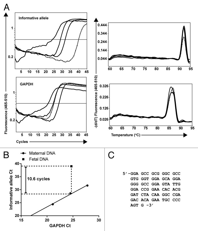
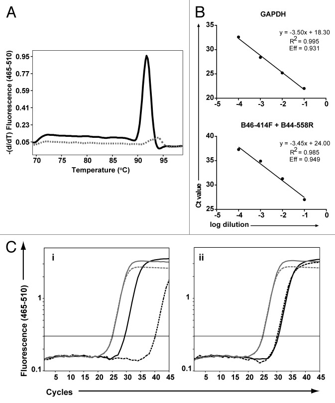
Although pregnancy-associated microchimerism is known to exist in humans, its clinical significance remains unclear. Fetal microchimerism has been documented in rhesus monkeys, but the trafficking and persistence of maternal cells in the monkey fetus and infant have not been fully explored. To investigate the frequency of maternal microchimerism in the rhesus monkey (Macaca mulatta), a real-time polymerase chain reaction (PCR) strategy was developed and validated to target polymorphic major histocompatibility complex (MHC) gene sequences. Informative PCR assays were identified for 19 of 25 dams and their respective offspring. Analyses were performed on tissues (thymus, liver, spleen, lymph nodes, and bone marrow) and peripheral blood mononuclear cells (PBMCs) collected prenatally and postnatally in a subset of animals. Seven of 19 monkeys had detectable maternal microchimerism in at least one compartment (range: 0.001-1.9% chimeric cells). In tissues, maternal microchimerism was found in 2 of 7 fetuses and 3 of 12 juveniles (1-1.5 years of age), and most of the animals that were positive had microchimeric cells in more than one tissue. Maternal microchimerism was detected in PBMCs from all (4 of 4) fetuses. These observations suggest that maternal microchimerism occurs in the rhesus monkey fetus and can be detected in tissues in a subset of offspring after birth.

Keywords: major histocompatibility complex; microchimerism; quantitative PCR; rhesus monkey; transplacental transfer.


Figures

None
Figure 1. Analysis of maternal microchimerism in fetal rhesus monkey samples using informative Mamu-MHC allele assays. (A) Three 10-fold serial dilutions of maternal DNA (solid lines) were amplified with the informative allele assay identified during the initial screening and with the GAPDH assay to control for genomic input. In parallel, a concentration of fetal DNA equivalent to that used in the intermediate diluted maternal sample (dashed line) was amplified using the same assays. The specificity of the maternal microchimeric signal detected in the fetal sample was verified by dissociation analysis (right panels). (B) The Ct values from amplification of the maternal dilutions with the informative allele and GAPDH assays (circles) were plotted to create a standard curve used to calculate the level of maternal microchimerism in the fetal sample (square). In the example presented, there is a 10.6 cycle difference between the informative allele signal detected in the fetal sample and the extrapolated signal for an equivalent genomic input of maternal sample. The calculated microchimerism level in the fetal sample is 2-10.6 = 0.06%. (C) The products amplified from maternal and fetal DNA were purified and sequenced using the amplification primers. A full-length sequence was obtained from both the maternal and fetal sample as shown. The sequence obtained from the fetal sample was identical to the sequence obtained from the maternal sample.
None
Figure 2. Performance of Mamu-MHC allele-specific real-time PCR assays used to identify microchimerism. (A) Samples from five different monkeys were amplified using A03–113F + A03–249R as a representative Mamu-MHC assay. Dissociation curves are shown for one animal positive for the target sequence (solid black curve) and one animal negative for the target sequence (dashed gray curve). (B) Four 10-fold serial dilutions of rhesus monkey DNA positive for the target sequence were spiked in PCR buffer (for GAPDH amplification) or in background rhesus monkey DNA negative for the target sequence (for MHC amplification). Standard curves are shown for GAPDH (used to control for total genomic input) and a representative Mamu-MHC assay. Amplification efficiency (Eff) was calculated based on the slope of the standard curve. (C) DNA from maternal-fetal monkey pairs was amplified with the assays shown in Table 2. Amplification curves are shown for GAPDH (in gray) to control for genomic input. Maternal samples are shown by solid lines and fetal samples are indicated by dashed lines. Examples of informative (Ci) and non-informative (Cii) allele assays for detection of maternal microchimerism are shown in black. The maternal sample is positive for the target sequence whereas the fetal sample may be positive for maternal microchimerism (Ci) or for the target sequence (Cii).

References

    1. Gammill HS, Nelson JL. Naturally acquired microchimerism. Int J Dev Biol. 2010;54:531–43. doi: 10.1387/ijdb.082767hg. - DOI - PMC - PubMed
    1. Hall JM, Lingenfelter P, Adams SL, Lasser D, Hansen JA, Bean MA. Detection of maternal cells in human umbilical cord blood using fluorescence in situ hybridization. Blood. 1995;86:2829–32. - PubMed
    1. Maloney S, Smith A, Furst DE, Myerson D, Rupert K, Evans PC, Nelson JL. Microchimerism of maternal origin persists into adult life. J Clin Invest. 1999;104:41–7. doi: 10.1172/JCI6611. - DOI - PMC - PubMed
    1. Saadai P, Lee TH, Bautista G, Gonzales KD, Nijagal A, Busch MP, Kim CJ, Romero R, Lee H, Hirose S, et al. Alterations in maternal-fetal cellular trafficking after fetal surgery. J Pediatr Surg. 2012;47:1089–94. doi: 10.1016/j.jpedsurg.2012.03.012. - DOI - PMC - PubMed
    1. Schröder J. Transplacental passage of blood cells. J Med Genet. 1975;12:230–42. doi: 10.1136/jmg.12.3.230. - DOI - PMC - PubMed

Publication types