. 2015 Jul;25(7):1815-26.
doi: 10.1093/cercor/bht419. Epub 2014 Jan 22.

The Rostro-Caudal Axis of Frontal Cortex Is Sensitive to the Domain of Stimulus Information

Affiliations

The Rostro-Caudal Axis of Frontal Cortex Is Sensitive to the Domain of Stimulus Information

Jörg Bahlmann et al. Cereb Cortex. 2015 Jul.

Abstract

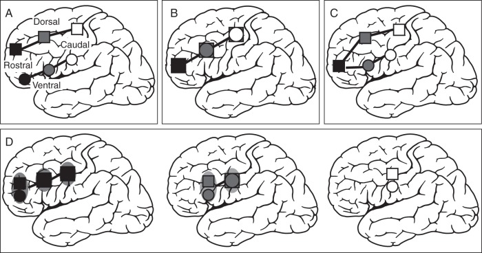
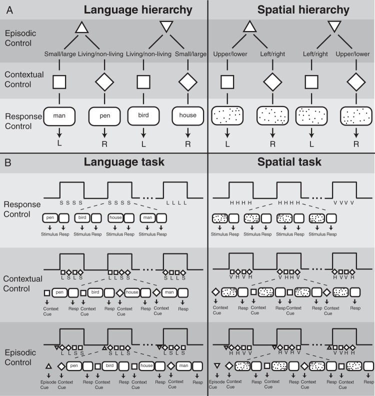
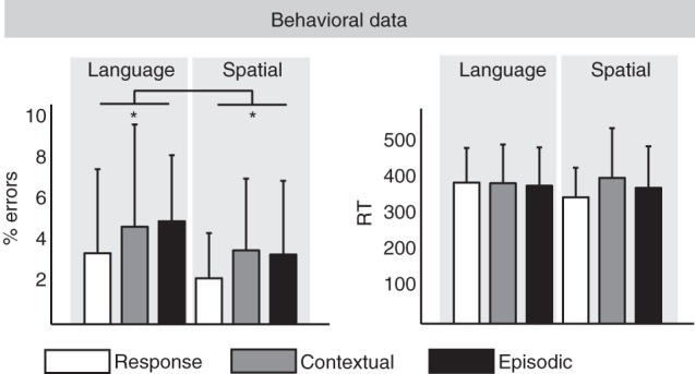
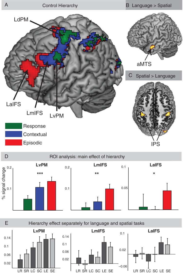
Evidence suggests that lateral frontal cortex implements cognitive control processing along its rostro-caudal axis, yet other evidence supports a dorsal-ventral functional organization for processes engaged by different stimulus domains (e.g., spatial vs. nonspatial). This functional magnetic resonance imaging study investigated whether separable dorsolateral and ventrolateral rostro-caudal gradients exist in humans, while participants performed tasks requiring cognitive control at 3 levels of abstraction with language or spatial stimuli. Abstraction was manipulated by using 3 different task sets that varied in relational complexity. Relational complexity refers to the process of manipulating the relationship between task components (e.g., to associate a particular cue with a task) and drawing inferences about that relationship. Tasks using different stimulus domains engaged distinct posterior regions, but within the lateral frontal cortex, we found evidence for a single rostro-caudal gradient that was organized according to the level of abstraction and was independent of processing of the stimulus domain. However, a pattern of dorsal/ventral segregation of processing engaged by domain-specific information was evident in each separable frontal region only within the most rostral region recruited by task demands. These results suggest that increasingly abstract information is represented in the frontal cortex along distinct rostro-caudal gradients that also segregate along a dorsal-ventral axis dependent on task demands.

Keywords: cognitive control; hierarchy; language; prefrontal cortex; spatial; stimulus domain.


Figures

Figure 1.
Figure 1.
Hypotheses tested in the present study. Cognitive control representations get more abstract from white to black. (A) Two rostro-caudal gradients of cognitive control separately for the 2 different stimulus domains (i.e., language–ventral, spatial–dorsal). (B) One rostro-caudal gradient of cognitive control. Overlapping activations for language and spatial domains along the rostro-caudal axis in lateral frontal cortex. (C) Lower levels of cognitive control recruit distinct ventral and dorsal subregions within caudal frontal cortex (e.g., language–ventral, spatial–dorsal). In contrast, more rostral regions of the lateral frontal cortex are independent of stimulus domain (i.e., overlapping activations for language and spatial stimuli in rostral regions). (D) Interaction between cognitive control and stimulus domain. Segregation of language (ventral) and spatial (dorsal) stimuli in subregions along the rostro-caudal axis, as a function of the cognitive control hierarchy. A stimulus domain segregation is only present at subregions that are engaged in the highest level of cognitive control.
Figure 2.
Figure 2.
(A) Schematic description of experimental design. In the Language Domain, participants made small/large or living/nonliving judgment of nouns. In the Spatial Domain, participants judged if a random dot pattern had more dots on the upper or lower part, or left or right part of the screen. Response Control: A stimulus (noun or dot pattern) was perceived and a response (button press) was made according to the type of task. Contextual Control: A cue (square or diamond) indicated which task to perform on each trial. Episodic Control: A cue (upward triangle vs downward triangle) indicated which subsequent cue (square or diamond) indicated which task to perform in a block of trials. (B) Schematic description of experimental tasks. One task block consisted of 4 consecutive trials. Each task block was separated by a resting baseline block of equal duration. Response Control: One task block comprised trials with identical tasks [e.g., Language: small/large (S) or living/nonliving (L) and Spatial: upper/lower (horizontal, H) or left/right (vertical, V) judgment]. Contextual Control: In each block, different tasks (Language: L or S and Spatial: H or V) were conducted based on a cue (square or diamond). Episodic Control: A cue–cue (upward triangle vs downward triangle) in front of each task block triggered the type cue–task association for the current block.
Figure 3.
Figure 3.
Behavioral results.
Figure 4.
Figure 4.
Patterns of activation across the whole brain for the (A) main effects of cognitive control. Each control condition collapsed across language and spatial tasks, (B) direct contrast of language versus spatial tasks collapsed across all cognitive control conditions, and (C) direct contrast of spatial versus language tasks collapsed across all cognitive control conditions. (D) Analysis of BOLD signal change in lateral frontal ROIs recruited by the cognitive control conditions collapsed across language and spatial tasks. A significant difference in signal change between the 3 different control tasks (Response, Contextual, and Episodic) was observed (*P < 0.05, **P < 0.005, ***P < 0.001). (E) Analysis in lateral frontal ROIs separately for each of the 6 conditions. LR, language task and response control; SR, spatial task and response control; LC, language task and contextual control; SC, spatial task and contextual control; LE, language task and episodic control; SE, spatial task and Episodic Control.
Figure 5.
Figure 5.
(A) Patterns of activation for the contrast of Episodic versus Contextual Control (red), Contextual versus Response Control (blue), and Response Control versus Baseline (green), P < 0.005, k = 30 voxels. (B) Patterns of activation for the 3 levels of cognitive control separately for language tasks. (C) Patterns of activation for the 3 levels of cognitive control separately for spatial tasks. (B,C) Activations for Response Control versus Baseline (green), Contextual Control versus Baseline (blue), and Episodic Control versus baseline (red) are shown with an uncorrected threshold of P < 0.001 and k = 30 adjacent voxels. Note that frontal activations in both plots do not survive multiple comparison corrections. See Figure 4A for (corrected) frontal activations of different levels of cognitive control collapsed across language and spatial tasks and see Figure 4B,C for the contrasts of language versus spatial, and vice versa collapsed across all cognitive control conditions.
Figure 6.
Figure 6.
Classification analysis results. (A) Distance between peak activation for language and spatial tasks on the z-axis. Bars represent mean distance values across participants in the 3 lateral frontal regions (LaIFS, LmIFS, and LvPM) for the cognitive control conditions (Epi, Episodic, Con, Contextual, and Res, Response; *P < 0.05; **P < 0.005). (B) Distribution of peak activity for language and spatial tasks in each participant is plotted in 3 lateral frontal regions for the cognitive control conditions. Individual MNI coordinates of language tasks (red triangles pointing down) and spatial tasks (blue triangles pointing up) were plotted in each ROI (overlapping language and spatial tasks are represented in overlapping triangles pointing down and pointing up). A significant dorsal–ventral segregation is illustrated with a green discrimination line. Lower left: Green discrimination line mapped on whole-brain activation pattern. Squares (light gray) represent bounding boxes of the search space for classification analysis. Bounding boxes were overlaid on activation pattern in lateral frontal regions using the MRIcron software (the coordinates of the bounding boxes are cartoon illustrations). (C) Distribution of the percentage of misclassification errors. A leave-one-participant-out cross-validation was applied on the classification analysis. The low percentage of misclassification errors represents better classification results and stronger dorsal–ventral topographical segregation in a given region and condition (*P < 0.001).

References

    1. Badre D, D'Esposito M. 2007. Functional magnetic resonance imaging evidence for a hierarchical organization of the prefrontal cortex. J Cogn Neurosci. 19:2082–2099. - PubMed
    1. Badre D, D'Esposito M. 2009. Is the rostro-caudal axis of the frontal lobe hierarchical? Nat Rev Neurosci. 10:659–669. - PMC - PubMed
    1. Badre D, Hoffman J, Cooney JW, D'Esposito M. 2009. Hierarchical cognitive control deficits following damage to the human frontal lobe. Nat Neurosci. 12:515–522. - PMC - PubMed
    1. Badre D, Poldrack RA, Paré-Blagoev EJ, Insler RZ, Wagner AD. 2005. Dissociable controlled retrieval and generalized selection mechanisms in ventrolateral prefrontal cortex. Neuron. 47:907–918. - PubMed
    1. Badre D, Wagner AD. 2007. Left ventrolateral prefrontal cortex and the cognitive control of memory. Neuropsychologia. 45:2883–2901. - PubMed

Publication types