. 2014 Jan 21;6(1):380-93.
doi: 10.3390/toxins6010380.

Active immunity induced by passive IgG post-exposure protection against ricin

Affiliations

Active immunity induced by passive IgG post-exposure protection against ricin

Charles Chen Hu et al. Toxins (Basel). .

Abstract

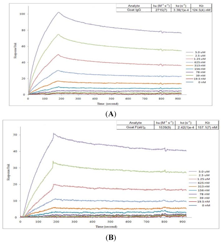
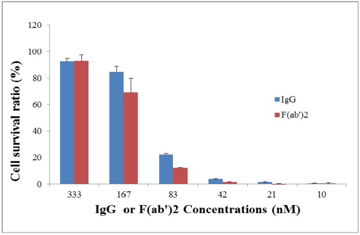
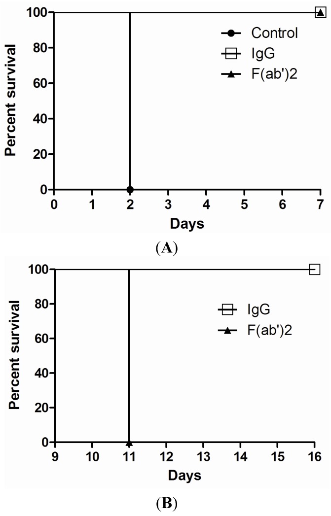
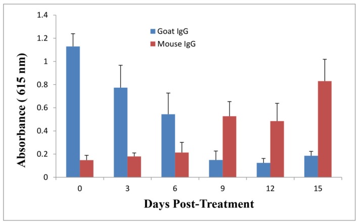
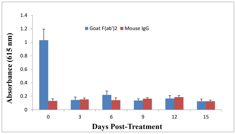
Therapeutic antibodies can confer an instant protection against biothreat agents when administered. In this study, intact IgG and F(ab')2 from goat anti-ricin hyperimmune sera were compared for the protection against lethal ricin mediated intoxication. Similar ricin-binding affinities and neutralizing activities in vitro were observed between IgG and F(ab')2 when compared at the same molar concentration. In a murine ricin intoxication model, both IgG and F(ab')2 could rescue 100% of the mice by one dose (3 nmol) administration of antibodies 1 hour after 5 × LD50 ricin challenge. Nine days later, when the rescued mice received a second ricin challenge (5 × LD50), only the IgG-treated mice survived; the F(ab')2-treated mice did not. The experimental design excluded the possibility of residual goat IgG responsible for the protection against the second ricin challenge. Results confirmed that the active immunity against ricin in mice was induced quickly following the passive delivery of a single dose of goat IgG post-exposure. Furthermore, it was demonstrated that the induced active immunity against ricin in mice lasted at least 5 months. Therefore, passive IgG therapy not only provides immediate protection to the victim after ricin exposure, but also elicits an active immunity against ricin that subsequently results in long term protection.


Figures

Figure 1
Figure 1
Sodium Dodecyl Sulfate Polyacrylamide Gel Electrophoresis (SDS-PAGE) analysis of goat F(ab’)2. Lane M is a molecular marker. Lanes 1 and 2 are goat IgG in non-reducing and reducing conditions. Lanes 3 and 4 are F(ab’)2 in non-reducing and reducing conditions.
Figure 2
Figure 2
In vitro binding affinity analysis for goat IgG and F(ab’)2 by Surface Plasmon Resonance (SPR). SPR sensorgram of the kinetics of association and dissociation of a range of concentrations from 0 to 5 µM of goat IgG (A) or F(ab’)2 (B) to immobilized ricin.
Figure 3
Figure 3
In vitro neutralization assay for goat IgG and F(ab’)2. Ricin (7.5 ng/mL) was pre-incubated with a serial dilution of goat IgG or F(ab’)2 for 2 h and then exposed to 104 Vero cells/well for 2 days before evaluation of cell viability using Alamar blue staining.
Figure 4
Figure 4
In vivo post-exposure protection assay for goat IgG and F(ab’)2. Ricin was given at the dose of 5 × LD50 to mice by i.p route. Goat IgG or F(ab’)2 at one dose of 3 nmol was administered i.p. at 1 h after ricin challenge and then mouse survival rate was monitored for 7 days (A); At day 9, mice were challenged with a second dose of 5 × LD50 of ricin and observed for 7 days (B). The experiment was repeated three times.
Figure 5
Figure 5
Evaluation of goat IgG and mouse IgG levels against ricin in pooled mouse sera over time by immunoassay. Goat IgG at the dose of 3 nmol was administered by the i.p. route into mice at 1 h post-challenge. Sera were collected at different time points for immunoassay to examine anti-ricin goat IgG and mouse IgG levels. All data represent the average of three independent experiments.
Figure 6
Figure 6
Evaluation of goat F(ab’)2 and mouse IgG levels against ricin in pooled mouse sera over time by immunoassay. Goat F(ab’)2 at the dose of 3 nmol was administered by the i.p. route into mice at 1 h post-challenge. Sera were collected at different time points for immunoassay to examine anti-ricin goat F(ab’)2 and mouse IgG levels. All data represent the average of three independent experiments.

References

    1. Zeitlin L., Cone R.A., Moench T.R., Whaley K.J. Preventing infectious disease with passive immunization. Microbes Infect. 2000;2:701–708. doi: 10.1016/S1286-4579(00)00355-5. - DOI - PubMed
    1. Little S.F., Ivins B.E., Fellows P.F., Friedlander A.M. Passive protection by polyclonal antibodies against bacillus anthracis infection in guinea pigs. Infect. Immun. 1997;65:5171–5175. - PMC - PubMed
    1. Casadevall A., Dadachova E., Pirofski L.A. Passive antibody therapy for infectious diseases. Nat. Rev. Microbiol. 2004;2:695–703. doi: 10.1038/nrmicro974. - DOI - PubMed
    1. Casadevall A. Passive antibody administration (immediate immunity) as a specific defense against biological weapons. Emerg. Infect. Dis. 2002;8:833–841. doi: 10.3201/eid0808.010516. - DOI - PMC - PubMed
    1. Casadevall A. Passive antibody therapies: Progress and continuing challenges. Clin. Immunol. 1999;93:5–15. doi: 10.1006/clim.1999.4768. - DOI - PubMed

Publication types