

Primary cardiac angiosarcoma - a review
- PMID: 24452054
- PMCID: PMC3907509
- DOI: 10.12659/MSM.889875
Primary cardiac angiosarcoma - a review
Abstract
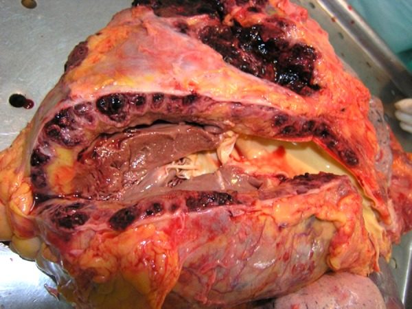
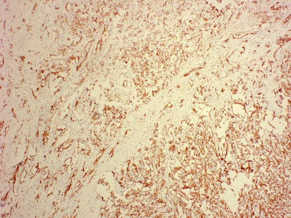
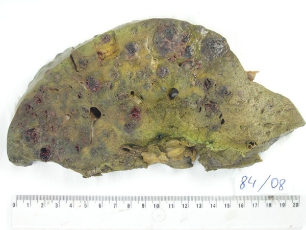
Primary cardiac neoplasms are extremely rare. Angiosarcoma is the most commonly seen histological subtype and is characterized by its permeating and destructive nature. Unfortunately, primary cardiac angiosarcoma is often overlooked as an initial diagnosis because of its rarity. Since the time it was first identified in 1934, little progress has been made in improving survival outcome. Complete or partial surgical resection is still the best option for palliation, with little hope for cure. Improvements have been made in the ability to view and distinguish tumors. Echocardiography is one of the most useful diagnostic tools because of its high sensitivity; therefore, CT and MR images are often used to detect sites of metastatic disease. Immunohistochemistry staining can also be employed as an adjunctive diagnostic tool. CD31, CD34, FLI-1, and von Willebrand factor are the most commonly used markers in detecting tumors of endothelial origin. However, due to the vast heterogeneity within a tumor, immunohistochemistry staining can be quite variable. Surgical resection remains the standard modality of treatment. Primary cardiac angiosarcoma is largely resistant to chemotherapy and/or radiation. However, the exact benefit and its place in a multimodality treatment regimen are still under investigation.
Figures
References
-
- Barnes AB, Beaver DC, Snell AM. Primary sarcoma of the heart: report of a case with electrocardiographic and pathological studies. Am Heart J. 2003;9:480–91.
-
- Look-Hong NP, Pandalai PK, Hornick JL, et al. Cardiac Angiosarcoma Management and Outcomes: 20-year single-institution experience. Ann Surg Oncol. 2012;19:2707–15. - PubMed
-
- Ferguson ER, Walsh GL. Sarcomas of the heart and great vessels. In: Pollock RE, editor. Soft tissue sarcomas. Hamilton: CB Decker Inc; 2002. pp. 155–56.pp. 158–60.
Publication types
MeSH terms
Substances
LinkOut - more resources
Full Text Sources
Other Literature Sources
