. 2014:5:3095.
doi: 10.1038/ncomms4095.

Actomyosin-dependent formation of the mechanosensitive talin-vinculin complex reinforces actin anchoring

Affiliations
Free PMC article

Actomyosin-dependent formation of the mechanosensitive talin-vinculin complex reinforces actin anchoring

Corina Ciobanasu et al. Nat Commun. 2014.
Free PMC article

Abstract

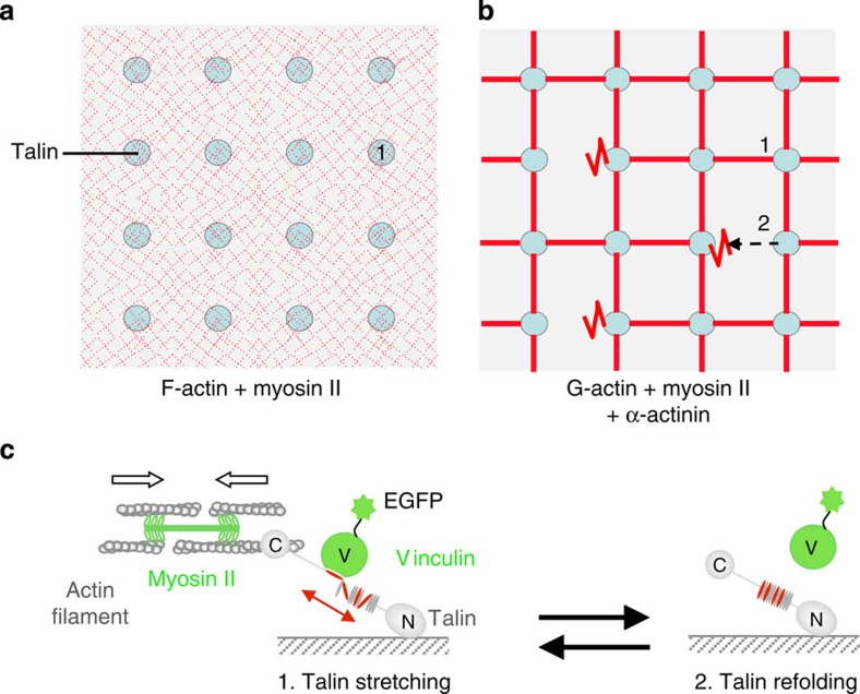
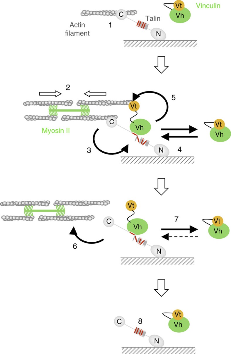
The force generated by the actomyosin cytoskeleton controls focal adhesion dynamics during cell migration. This process is thought to involve the mechanical unfolding of talin to expose cryptic vinculin-binding sites. However, the ability of the actomyosin cytoskeleton to directly control the formation of a talin-vinculin complex and the resulting activity of the complex are not known. Here we develop a microscopy assay with pure proteins in which the self-assembly of actomyosin cables controls the association of vinculin to a talin-micropatterned surface in a reversible manner. Quantifications indicate that talin refolding is limited by vinculin dissociation and modulated by the actomyosin network stability. Finally, we show that the activation of vinculin by stretched talin induces a positive feedback that reinforces the actin-talin-vinculin association. This in vitro reconstitution reveals the mechanism by which a key molecular switch senses and controls the connection between adhesion complexes and the actomyosin cytoskeleton.


Figures

Figure 1
Figure 1. Experimental strategies used in this study.
(a) In this assay, a preformed actomyosin network (red), made of short actin filaments (F-actin) and myosin II filaments, generates force on talin-containing disc-shaped islands micropatterned on a glass coverslip. The force generated by this actomyosin network is sufficient to stretch talin and induce vinculin binding (see 1 in panel c). This experimental setup favours an instantaneous, homogeneous and synchronous mechanosensitive response. (b) Monomeric actin (G-actin), myosin II and α-actinin self-assemble into dynamic actomyosin cables (red) that control the association of vinculin to talin in a reversible manner. In these conditions, actomyosin cables stretch talin, allowing vinculin binding (see 1 in the panel c). The detachment of the cables allows vinculin dissociation and talin refolding (see 2 in the panel c). (c) Details of the reactions mentioned in (a) and (b). The mechanosensitive domain of talin corresponds to a group of helix bundles in the talin rod (amino acids 482–889) described in refs , . In talin, the vinculin-binding sites (VBSs) are in red and the other helices are in grey.
Figure 2
Figure 2. In vitro reconstitution of the actomyosin-dependent binding of vinculin to talin.
(af) Recruitment of Vh to talin-coated discs in the presence of an actomyosin network. Conditions: 22 nM of EGFP-Vh, 0.12 μM Myosin II, 2.4 μM of short actin filaments capped at their barbed end (2% Alexa594-labelled). The binding of EGFP-Vh and actin to talin-coated discs is shown at steady state in the presence of both actin and myosin (a,d), actin alone (b,e) and myosin alone (c). The pictures show EGFP-Vh (ac) and actin (d,e). Scale bar, 15 μm. Supplementary Movie 1. (f) Kymographs of the cross-sections drawn in (ae). Scale bar, 6 μm (horizontal), 300 s (vertical). (g) Kinetics of EGFP-Vh recruitment to talin-coated discs in the conditions described in the previous panels (af). (h) Steady-state fluorescence of talin-bound EGFP-Vh as a function of EGFP-Vh concentration in the presence of both actin and myosin or myosin alone as described in (af). Data are mean±s.d., n≥8. Note that the experiment presented in (g) has been acquired with different camera settings and the fluorescence values cannot be compared with (h). (i) Maximum rate (Vmax) of EGFP-Vh binding to talin-coated discs as a function of EGFP-Vh concentration in the presence of actin and myosin as described in (af). Data are mean±s.d., n≥8. (j) Maximum rate (Vmax) of EGFP-Vh binding to talin-coated discs, in the conditions described in (af), as a function of myosin concentration. Data are mean±s.d., n≥8. (hj), data collected in two independent experiments.
Figure 3
Figure 3. α-actinin controls the stability of the actomyosin cables.
(a) Self-assembly of actomyosin cables associated with talin-coated discs. Conditions: 2.4 μM G-actin (2% Alexa594-labelled), 0.12 μM Myosin II and 62 nM of α-actinin. Scale bar, 35 μm. Supplementary Movie 2. (b) Time lapse showing the formation of a cable between two talin-coated discs extracted from (a). Scale bar, 35 μm. (c) Self-assembly of actomyosin cables associated with talin-coated discs. Conditions: 2.4 μM G-actin, 0.12 μM Myosin II and the indicated concentrations of mCherry-α-actinin. Scale bar, 35 μm. Supplementary Movie 3. (d) Kymographs showing the retraction of mCherry-α-actinin-containing cables, at the indicated concentrations of mCherry-α-actinin. Scale bar, 10 μm (horizontal), 100 s (vertical). (e) Quantification of the retraction speed as a function of the fluorescence intensity of mCherry-α-actinin along the detaching cables, at the indicated concentrations of mCherry-α-actinin. (f) Quantification of the number of cables (in pictures showing 8 discs) and the frequency of detachment of the cables during time intervals of 400 s (in movies showing 8 discs) as a function of the concentration of mCherry-α-actinin. Data are mean±s.d. Number of cables n≥20, frequency of detachment n=6. Data collected in two independent experiments. The conditions for (df) are described in (c).
Figure 4
Figure 4. Parameters that control the lifetime of the talin–vinculin complex.
(a) Time lapse showing the colocalization of actomyosin cables and EGFP-Vh associated with talin-coated discs. Conditions: 22 nM of EGFP-Vh, 0.12 μM Myosin II, 2.4 μM of G-actin (2% Alexa594-labelled) and 0.12 μM α-actinin. Scale bar, 15 μm. Supplementary Movie 4. (b) Kymographs showing the detachment of the cable marked with an arrowhead in (a), followed by the dissociation of EGFP-Vh. Scale bar, 10 μm (vertical), 300 s (horizontal). (c) Quantification of the kymographs presented in (b). (d) Comparison between the kinetics of dissociation of EGFP-Vh from talin measured by FRAP when a constant force is applied (Supplementary Fig. 3c,d, Supplementary Movie 5) and after the detachment of a cable (conditions described in Fig. 4a). Data were normalized between 1 and 0 to be compared. The inset shows a quantification of the dissociation rates. Data are mean±s.d. and the P-value was calculated (P=0.52) using a t-test. Dissociation after force release, n=12; dissociation during force application, n=28. Data collected in three independent experiments. (e) Maximum intensity of EGFP-Vh fluorescence reached in the discs as a function of α-actinin concentration. Data are mean±s.d., n≥32. Data collected in three independent experiments. (f) Distribution of the lifetime (time during which the intensity is 60% of the maximum) of EGFP-Vh spots associated with actomyosin cables as a function of α-actinin concentration. For (e) and (f), the conditions were described in (a). NS, not significant.
Figure 5
Figure 5. A positive feedback stabilizes the actomyosin–talin–vinculin association.
(a) Self-organization of actomyosin structures between talin-coated discs in the presence of EGFP-Vh and EGFP-vinculin as indicated. Conditions: 2.2 μM of EGFP-vinculin (or EGFP-Vh), 0.4 μM Myosin II, 1.2 μM of G-actin (2% Alexa594-labelled). In this series of experiments the discs are coated with a low concentration of talin (0.1 μM in the coating reaction) to make the anchoring of the cable a limiting parameter. Scale bar, 15 μm. Supplementary Movie 6. (b,c) Representative kinetics of EGFP-Vh/actin (b) and EGFP-vinculin/actin (c) binding to talin-coated discs. The regions of interest are indicated in (a). (d) Quantification of the rate at which actin is recruited to talin-coated discs after EGFP-Vh and EGFP-vinculin reached steady state (from 1,500 s to 2,200 s). Data are mean±s.d. For both EGFP-Vh and EGFP-vinculin, n=32 regions of interest collected in two independent experiments. The P-value was calculated (P<0.0001) using a t-test.
Figure 6
Figure 6. Model for the actomyosin-dependent dynamics of the talin–vinulin complex.
The mechanosensitive domain of talin is described at the end of the legend of Fig. 1. (1) Actin filament capture by talin. (2) Self-assembly of actomyosin cables. (3) Talin stretching, VBSs exposure. (4) Association and activation of vinculin. (5) Vinculin binding to actin and reinforcement of the anchoring. (6) Detachment of the cable. (7) Dissociation of vinculin. (8) Talin refolding, no reassociation of vinculin.

References

    1. Wehrle-Haller B. Structure and function of focal adhesions. Curr. Opin. Cell Biol. 24, 116–124 (2012). - PubMed
    1. Gardel M. L., Schneider I. C., Aratyn-Schaus Y. & Waterman C. M. Mechanical integration of actin and adhesion dynamics in cell migration. Annu. Rev. Cell Dev. Biol. 26, 315–333 (2010). - PMC - PubMed
    1. Zaidel-Bar R., Ballestrem C., Kam Z. & Geiger B. Early molecular events in the assembly of matrix adhesions at the leading edge of migrating cells. J. Cell Sci. 116, 4605–4613 (2003). - PubMed
    1. Kuo J. C., Han X., Hsiao C. T., Yates J. R. 3rd & Waterman C. M. Analysis of the myosin-II-responsive focal adhesion proteome reveals a role for beta-Pix in negative regulation of focal adhesion maturation. Nat. Cell Biol. 13, 383–393 (2011). - PMC - PubMed
    1. Geiger B., Spatz J. P. & Bershadsky A. D. Environmental sensing through focal adhesions. Nat. Rev. Mol. Cell Biol. 10, 21–33 (2009). - PubMed

Publication types