. 2014 Jan 23:4:3838.
doi: 10.1038/srep03838.

Characterization of cleavage intermediate and star sites of RM.Tth111II

Affiliations

Characterization of cleavage intermediate and star sites of RM.Tth111II

Zhenyu Zhu et al. Sci Rep. .

Abstract

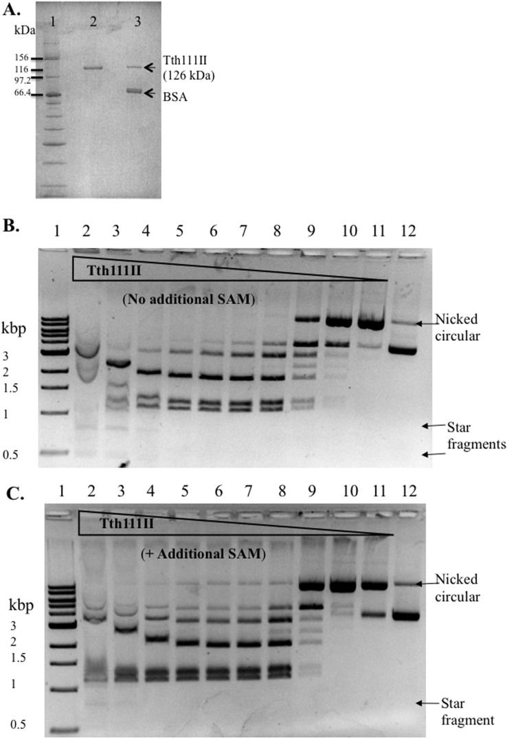
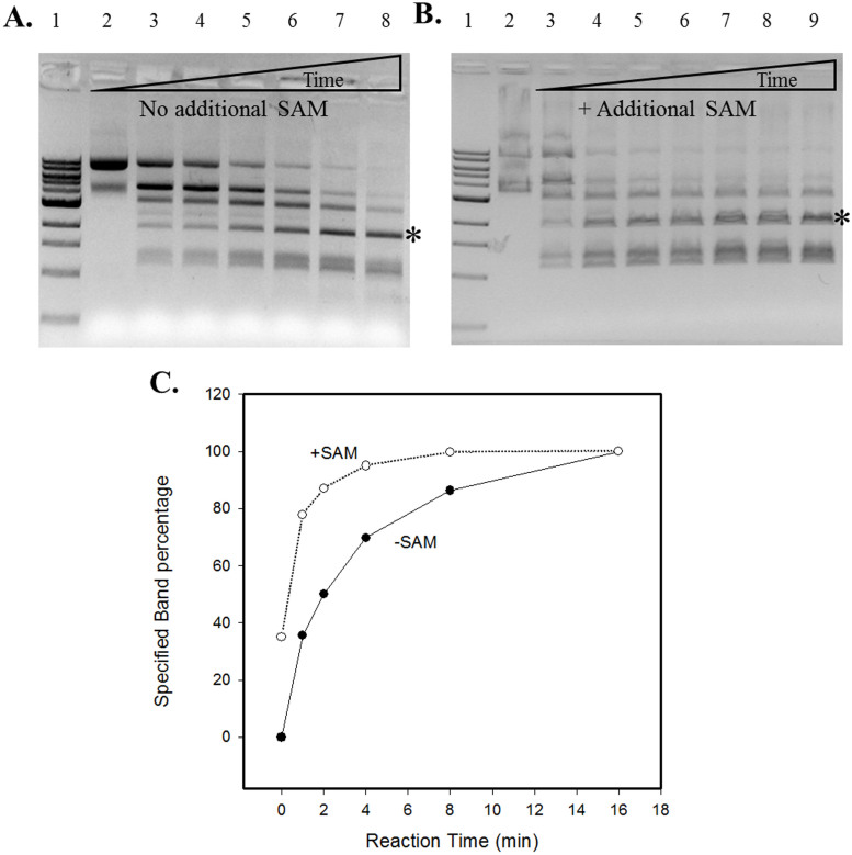
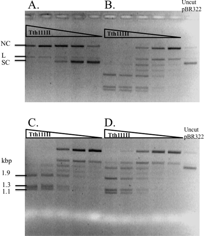
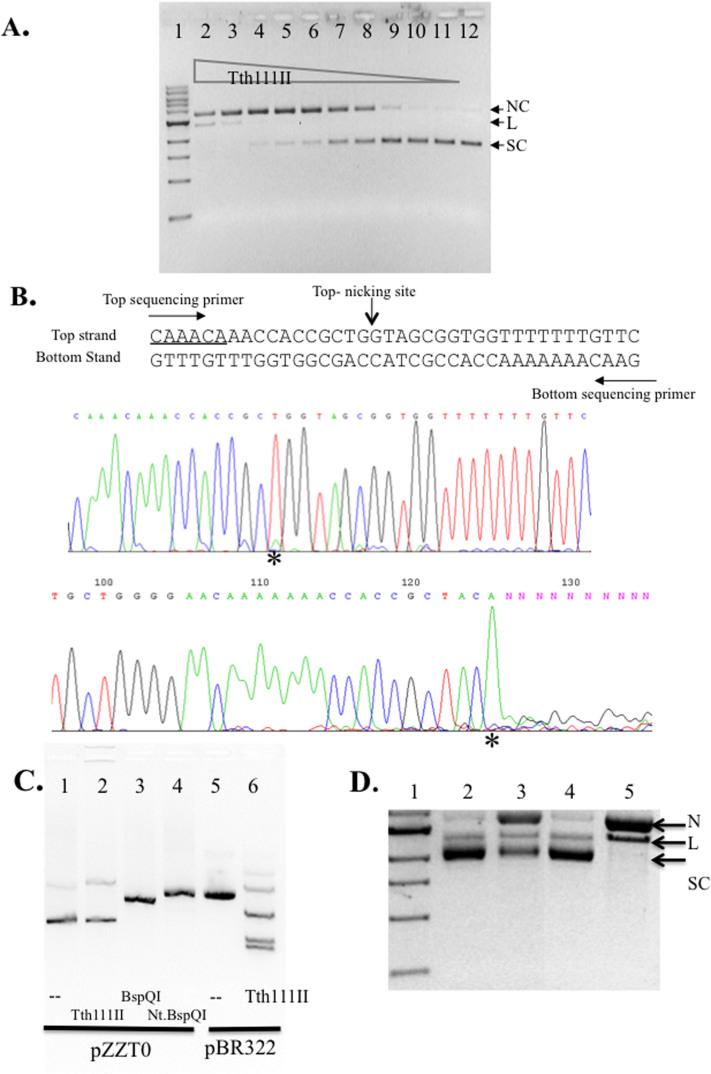
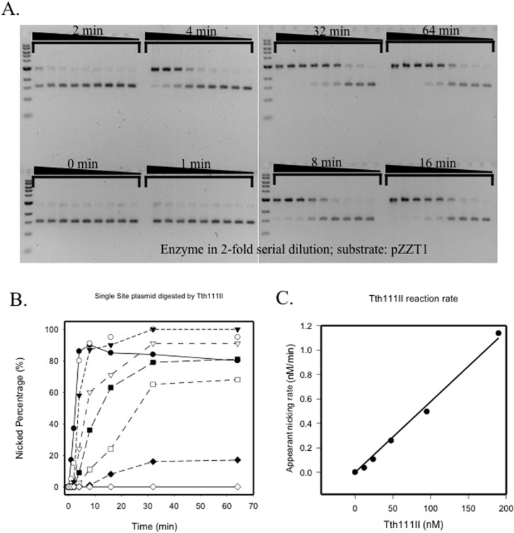
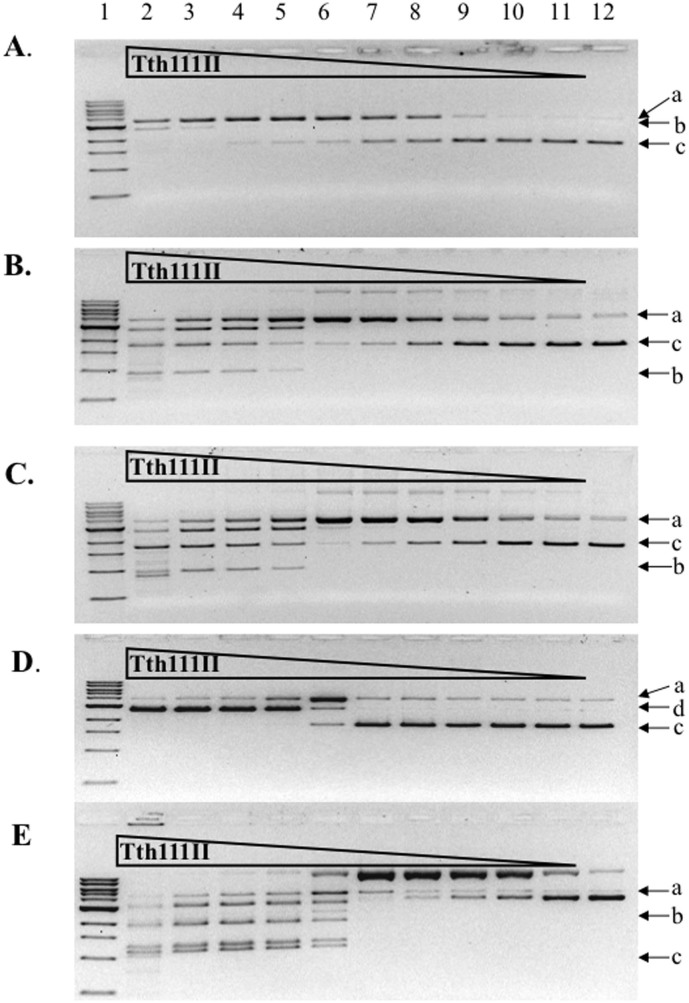
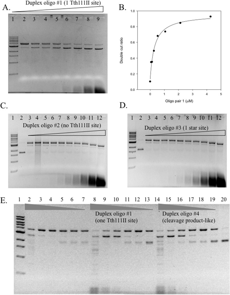
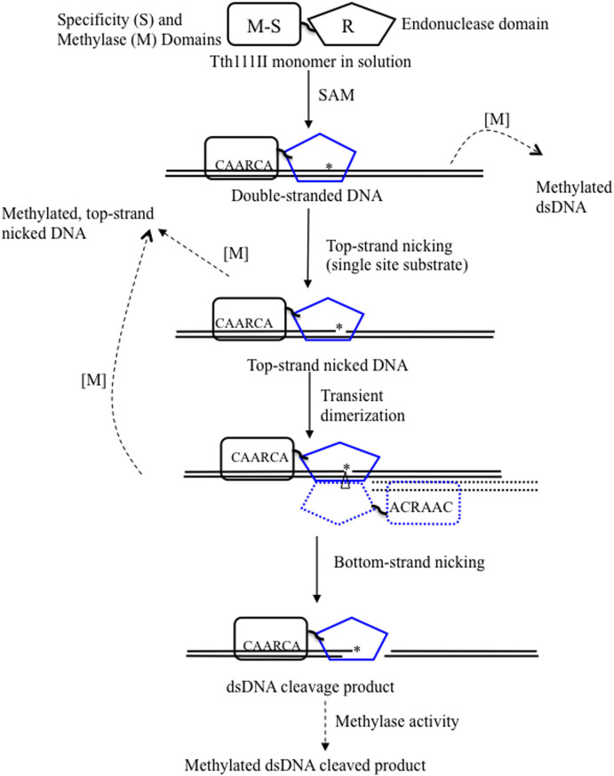
Tth111II is a thermostable Type IIGS restriction enzyme that recognizes DNA sites CAARCA (R = A or G) and cleaves downstream at N11/N9. Here, the tth111IIRM gene was cloned and expressed in E. coli, and Tth111II was purified. The purified enzyme contains internally-bound S-adenosylmethionine (SAM). When the internal SAM was removed, the endonuclease activity was stimulated by adding SAM or its analog sinefungin. The cleavage intermediate is mostly top-strand nicked DNA on a single-site plasmid. Addition of duplex oligos with a cognate site stimulates cleavage activity of the one-site substrate. Tth111II cleaves a two-site plasmid DNA with equal efficiency regardless of site orientation. We propose the top-strand nicking is carried out by a Tth111II monomer and bottom-strand cleavage is carried out by a transient dimer. Tth111II methylates cleavage product-like duplex oligos CAAACAN9, but the modification rate is estimated to be much slower than the top-strand nicking rate. We cloned and sequenced a number of Tth111II star sites which are 1-bp different from the cognate sites. A biochemical pathway is proposed for the restriction and methylation activities of Tth111II.


Figures

Figure 1
Figure 1. SDS-PAGE analysis of recombinant and native Tth111II, and endonuclease activity assays.
(A). Lane 1, protein size marker; Lane 2, purified recombinant Tth111II; lane 3, purified native Tth111II plus BSA. (B). Tth111II endonuclease activity in the absence of additional SAM. Lanes 1, 1 kb DNA marker. Lanes 2–11: 2-fold serial dilution of Tth111II in digestion of 0.5 μg pBR322 (16 units (365 pmole) in lane 2); lane 12, uncut pBR322. Reactions were carried out in Tth Buffer at 65°C for 30 min. (C). Tth111II endonuclease activity in the presence of additional 320 μM SAM.
Figure 2
Figure 2. Time course study of Tth111II digestion in the absence or presence of additional SAM.
The reaction conditions were same as described in Figure 1 legend, but with 4 units per lane for various times. Panel (A): no additional SAM added. Lane 1, 1 kb DNA ladder; Lane 2–8: 0, 1, 2, 4, 8, 16, 32 min digestion. Panel (B), 320 μM SAM added; lane 2, uncut pBR322; lanes 3–9: 0, 1, 2, 4, 8, 16, 32 min digestion. (C): A graphic representation of one cleavage product band indicated by “*” as the result of Tth111II digestion with or without additional SAM.
Figure 3
Figure 3. Effects of SAM and sinefungin cofactor on SAM-depleted Tth111II endonuclease activity.
Panel (A), SAM-depleted Tth111II digestion of 1 μg pBR322. Panel (B), same as in (A), but with addition of 320 μM SAM. Panel (C), same as in (A), plus 1 mM sinefungin. (D), untreated Tth111II with internally bound SAM. Reaction was carried out in Tth Buffer, 65°C for 30 min. The triangle on top of lanes represents a 2-fold serial dilution of the enzyme (starting at 11.4 pmole). NC, nicked circular; L, linear, SC, supercoiled DNA.
Figure 4
Figure 4. Tth111II activity assay on a single-site substrate and run-off sequencing of the nicked products.
(A). Tth111II digestion of pZZT1 with a single Tth111II site plus 320 μM SAM. Lane 2–11; 220 ng pZZT1 was digested with Tth111II in a 2-fold enzyme serial dilution (lane 2 enzyme = 91 pmole); Lane 12, uncut pZZT1. (B). Run-off sequencing of the nicked pZZT1. The template sequence and the sequencing primers and nicking sites are shown. Top sequencing panel, sequence read from the top sequencing primer. Bottom sequencing panel, sequence read from bottom sequencing primer. “*” indicates extra “A” peak in the run-off reactions. (C). Digestion of pZZT0 (no Tth111II site) by Tth111II endonuclease. (D). Stepwise methylase and endonuclease activity assays for RM.Tth111II in the absence of or presence of Mg2+. Lane 1, DNA marker; lanes 2, partially modified pZZT1 after methylation in the digestion buffer in the absence of Mg2+; lane 3, endonuclease digestion of the partially modified pZZT1 in the presence of Mg2+ and SAM (note, the remaining supercoiled DNA is resistant to digestion/nicking due to the methylation of Tth111II site. Thus the remaining supercoiled DNA is an indication of methylase activity of RM.Tth111II); lane 4, Mock-modified (unmethylated) pZZT1; lane 5, mock-modified pZZT1 DNA digested by Tth111II in the presence of Mg2+. N, nicked circular; L, linear DNA; S, supercoiled DNA.
Figure 5
Figure 5. Kinetics study of Tth111II top-strand nicking.
(A). pZZT1 digested by Tth111II (+320 μM SAM) in 2-fold serial dilutions of the enzyme and in different times: 0, 1, 2, 4, 8, 16, 32 and 64 min. (B). Graphic representation of the nicked circular DNA as percentage of total DNA in the time course. (C). The linear relationship of the apparent nicking rate with Tth111II enzyme concentration.
Figure 6
Figure 6. Tth111II digestion of five plasmid substrates in the presence of 320 μM SAM.
(A). Tth111II digestion of 220 ng pZZT1 with a single site. (B). Tth111II digestion of 500 ng pZZ2TF with two sites in the same orientation. (C). Tth111II digestion of 500 ng pZZ2TR with two sites in the reverse orientation. (D). Tth111II digestion of 500 ng pUC19 with three closely located sites. (E). Tth111II digestion of 500 ng pBR322 with five sites. (A), open circular nicked DNA; (b), dsDNA cut product; (c), supercoiled DNA; (d), linear DNA from double-strand cuts or two sites with nicks on opposite strands. Enzyme in lane 2 of ABCDE = 91 pmole.
Figure 7
Figure 7. Oligonucleotide stimulation of dsDNA cleavage activity.
(A). Tth111II digestion of pZZTB1 (Nb.BsmI pre-nicked) with the addition of different concentrations of oligos in 30 min at 65°C (+320 μM SAM). Lane 1, 1 kb DNA ladder; Lane 2–9; Tth111II digestion of pZZTB1 (Nb.BsmI pre-nicked) in the presence of 0, 0.067, 0.13, 0.27, 0.53, 1.1, 2.1, 4.3 μM of duplex oligo 1 (one Tth111II site). The final cleavage product is linear DNA. (B). Graphic representation of the relationship of dsDNA cut ratio and concentration of the duplex oligo 1 as shown in panel A. (C). Same reaction condition as in A. Lane 2, uncut pZZTB1; lane 3–12: Tth111II digestion of pZZTB1 (Nb.BsmI pre-nicked) in the presence of 0.017, 0.033, 0.067, 0.13, 0.27, 0.53, 1.1, 2.1, 4.3 μM of duplex oligos 2 (no Tth111II site). (D). Same as in C except the duplex oligos is #3 with one Tth111II star site. (E). Same reaction condition as in A. Lane 2–7, Tth111II digestion of pZZT1 by decreasing amount of Tth111II enzyme without additional duplex oligos. Lane 8–13, Tth111II digestion of pZZTB1 by decreasing amount of Tth111II enzyme plus duplex oligos 1. Lane 14–19, same as lanes 8–13, except the duplex oligo is #4 (cleavage product-like oligos).
Figure 8
Figure 8. Possible reaction steps for RM.Tth111II endonuclease and methylase activities.
“*” indicates top-strand (CAARCA) nicking; “Δ” indicates bottom-strand nicking; solid arrows, endonuclease activity; dashed arrow, methylase activity; solid blocks: Tth111II enzyme; solid line, target DNA; dotted block and dotted line, a second enzyme molecule which may be bound to duplex oligos with one site or cleavage product oligos with a cognate site. Blue color endonuclease domain indicates possible conformation changes after binding to the cognate site and SAM after allosteric activation.

Similar articles

Cited by

References

    1. Roberts R. J., Vincze T., Posfai J. & Macelis D. REBASE--a database for DNA restriction and modification: enzymes, genes and genomes. Nucleic Acids Res 38, D234–236 (2010). - PMC - PubMed
    1. Roberts R. J. et al. A nomenclature for restriction enzymes, DNA methyltransferases, homing endonucleases and their genes. Nucleic Acids Res 31, 1805–1812 (2003). - PMC - PubMed
    1. Matvienko N. N., Kramarov V. M., Ivanov L. & Matvienko N. I. Bce83I, a restriction endonuclease from Bacillus cereus 83 which recognizes novel nonpalindromic sequence 5′-CTTGAG-3′ and is stimulated by S-adenosylmethionine. Nucleic Acids Res 20, 1803 (1992). - PMC - PubMed
    1. Shapovalova N. I., Zheleznaia L. A., Matvienko N. N. & Matvienko N. I. [A new site-specific endonuclease BspKT51]. Biokhimiia 59, 485–493 (1994). - PubMed
    1. Janulaitis A., Bitinaite J. & Jaskeleviciene B. A new sequence-specific endonuclease from Gluconobacter suboxydans. FEBS Lett 151, 243–247 (1983). - PubMed

Publication types

MeSH terms

LinkOut - more resources