Review
. 2014 Feb;15(2):108-21.
doi: 10.1038/nrm3742.

A day in the life of the spliceosome

Affiliations
Review

A day in the life of the spliceosome

A Gregory Matera et al. Nat Rev Mol Cell Biol. 2014 Feb.

Erratum in

  • Nat Rev Mol Cell Biol. 2014 Apr;15(4):294

Abstract

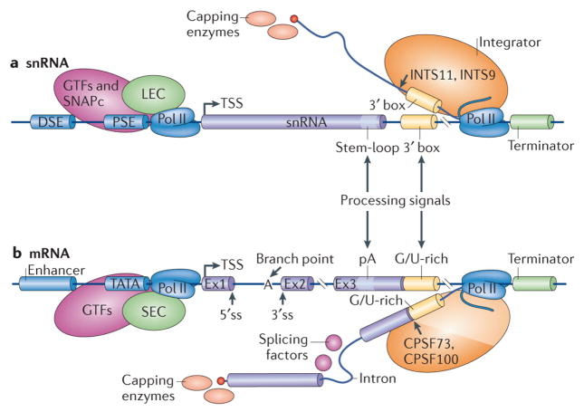
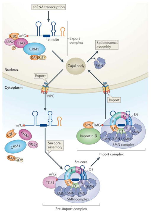
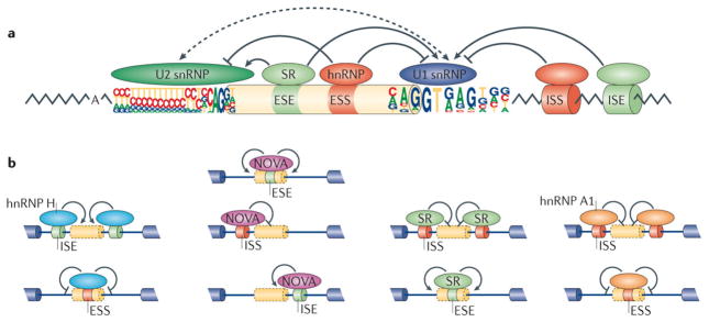
One of the most amazing findings in molecular biology was the discovery that eukaryotic genes are discontinuous, with coding DNA being interrupted by stretches of non-coding sequence. The subsequent realization that the intervening regions are removed from pre-mRNA transcripts via the activity of a common set of small nuclear RNAs (snRNAs), which assemble together with associated proteins into a complex known as the spliceosome, was equally surprising. How do cells coordinate the assembly of this molecular machine? And how does the spliceosome accurately recognize exons and introns to carry out the splicing reaction? Insights into these questions have been gained by studying the life cycle of spliceosomal snRNAs from their transcription, nuclear export and re-import to their dynamic assembly into the spliceosome. This assembly process can also affect the regulation of alternative splicing and has implications for human disease.


Figures

Figure 1
Figure 1. Comparison of transcription and processing of snRNAs and mRNAs
Sm-class snRNA genes (a) share a number of common features with protein-coding mRNA genes (b), including the arrangement of upstream and downstream control elements. The cis-acting elements and trans-acting factors involved in expression of these two types of transcripts are depicted. The DSE (distal sequence element) and PSE (proximal sequence element) are roughly equivalent to the enhancer and TATA box elements, respectively, of mRNA genes. Positive transcription elongation factor b (P-TEFb; not shown) is recruited to both promoters by RNA pol II. In addition, snRNA promoters recruit the LEC (little elongation complex), whereas mRNA promoters recruit the SEC (super elongation complex). Initiation of snRNA transcription requires general transcription factors (GTFs) as well as the snRNA-activating protein complex (SNAPc). The Integrator complex is required for recognition of snRNA downstream processing signals, including the 3′ box. Two of its subunits, IntS11 and IntS9, share sequence similarity to the mRNA 3′ processing factors CPSF73 and CPSF100. For both snRNAs and mRNAs, 5′ end capping and 3′ end cleavage are thought to occur co-transcriptionally. Additional processing factors (not shown) are recruited to the nascent transcripts via interactions with the pol II C-terminal domain.
Figure 2
Figure 2. Maturation of snRNAs requires nuclear and cytoplasmic regulatory steps
The snRNA pre-export complex consists of the heterodimeric cap-binding complex (CBC), arsenite resistance protein 2 (ARS2), the hyperphosphorylated form of the export adaptor PHAX and the large multi-subunit Integrator complex (not shown). Upon release from the site of snRNA transcription, the pre-export complex is remodelled within the nucleoplasm to form the export complex. This step involves removal of Integrator proteins and binding of the export receptor CRM1 (chromosome region maintenance 1) and the GTP-bound form of the RAN GTPase. Nucleoplasmic remodelling probably includes a Cajal body-mediated surveillance step to ensure RNP quality. Once transported to the cytoplasm, these export factors dissociate from the pre-snRNA prior to Sm core assembly and exonucleolytic trimming of the snRNA 3′ end (orange stem sloop). Following assembly of the Sm core snRNP (detailed in Fig. 3), the 7-methylguanosine (m7G) cap is hypermethylated by TGS1 (trimethylguanosine synthase 1) to form a 2,2,7-trimethylguanosine (TMG) cap. Generation of the TMG cap triggers assembly of the import complex, which includes the import adaptor snurportin (SPN) and the import receptor importin-β; both SPN and the SMN complex make functional contacts with importin-β (for simplicity, other components of the SMN complex are not depicted). Upon nuclear re-entry, the Sm snRNPs transiently localize to Cajal bodies for nuclear maturation steps, followed by dissociation from SMN and storage within splicing factor compartments called nuclear speckles. Spliceosome assembly (detailed in Fig. 4) takes place at sites of pre-mRNA transcription.
Figure 3
Figure 3. Assisted assembly of Sm-class snRNPs
Following their translation, Sm proteins are sequestered and symmetrically dimethylated by the PRMT5 complex. Once formed, the 6S complex of the Sm (D1-D2-F-E-G) pentamer and pICln is thought to be released from PRMT5c as a separate particle. This 6S complex is delivered to the oligomeric, multi-subunit SMN complex, which provides the overall platform for subsequent assembly steps. Gemin2, (Gem2), the heterodimeric binding partner of SMN, binds to the 6S complex, forming an early 8S assembly intermediate. In parallel, the SMN complex, including Gemin5 (Gem5), recognizes specific sequence elements (the Sm-site and the 3′ stem-loop) within the post-export snRNA. A poorly understood series of rearrangements leads to formation of the assembled core snRNP. These involve recruitment of the m7G-capped snRNA and the SmB-SmD3-pICln subcomplex, followed by dissociation of pICln. Prior to SmB-SmD3 incorporation, the ‘horseshoe’ intermediate may be stabilized by the Tudor domain of SMN, which contains an Sm fold. Incorporation of SmB-SmD3 and completion of the heteroheptameric ring requires the presence of an RNA that contains an Sm site. This produces an assembled core snRNP that is ready for downstream events including TMG capping and formation of the nuclear import complex (see Fig. 2).
Figure 4
Figure 4. Step-wise assembly of the spliceosome and catalytic steps of splicing
Spliceosome assembly takes place at sites of transcription. (a) The U1 and U2 snRNPs assemble onto the pre-mRNA in a co-transcriptional manner through recognition of the 5′ and 3′ splice sites, which is mediated by the C-terminal domain (CTD) of pol II. The U1 and U2 snRNPs interact with each other to form the pre-spliceosome (complex A). This process is dependent on DExD/H helicases Prp5 and Sub2. In a subsequent reaction catalysed by Prp28, the preassembled tri-snRNP U4/U6•U5 is recruited to form complex B. The resulting complex B undergoes a series of rearrangements to form a catalytically active complex B (complex B*), which requires multiple RNA helicases (Brr2, Snu114 and Prp2) and results in the release of U4 and U1 snRNPs. Complex B* then carries out the first catalytic step of splicing, generating complex C, which contains the free exon 1 and the intron-exon 2 lariat intermediate. Complex C undergoes additional rearrangements and then carries out the second catalytic step, resulting in a post-spliceosomal complex that contains the lariat intron and spliced exons. Finally, the U2, U5 and U6 snRNPs are released from the mRNP particle and recycled for additional rounds of splicing. Release of the spliced product from the spliceosome is catalysed by the DExD/H helicase Prp22, . (b) During splicing, RNA-RNA interactions are rearranged in a stepwise manner to create the catalytic center of the spliceosome. Initially, U1 and U2 snRNA pair with the 5′ss and the branch point sequence within complex A (left, the branch point adenosine is indicated). Subsequently, complex A associates with the U4/U6•U5 tri-snRNP, leading to new base pairs between U2 and U6 snRNA and between U5 snRNA and exonic sequences near the 5′ss (middle). The U4 snRNA is disassociated from U6 to expose the 5′ end of U6, which then base pairs with the 5′ss to displace U1 snRNA (right). In the end, an extensive network of base pairing interactions is formed between U6 and U2, juxtaposing the 5′ss and branch point adenosine for the first catalytic step of splicing. The central region of U6 snRNA forms an intramolecular stem-loop (the U6-ISL) that is key for splicing catalysis.
Figure 5
Figure 5. Regulation of alternative splicing
(a) Splice site choice is regulated through cis-acting splicing regulatory elements (SREs) and trans-acting splicing factors. Based on their relative locations and activities, SREs can be classified as exonic or intronic splicing enhancers and silencers (ESEs, ISEs, ESSs or ISSs). These SREs specifically recruit splicing factors to promote or inhibit recognition of nearby splice sites. Common splicing factors include SR proteins that recognize ESEs to promote splicing, as well as various hnRNPs that typically recognize ESSs to inhibit splicing. Both often affect the function of U2 and U1 snRNPs during spliceosomal assembly. (b) The activity of splicing factors and cis-acting SREs is context-dependent. Four well characterized examples are shown from left to right. Oligo-G tracts, recognized by hnRNP H, function as ISEs to promote splicing when they are located inside an intron or as ESSs when located within exons (left). YCAY motifs, recognized by NOVA, act as ESEs when located inside an exon, as ISSs when located in the upstream intron of an alternative exon, or as ISEs when located in the downstream intron. Binding sites for SR proteins or hnRNP A1 also have distinct activities when located at different regions on the pre-mRNA.

References

    1. Berget SM, Moore C, Sharp PA. Spliced segments at the 5′ terminus of adenovirus 2 late mRNA. Proceedings of the National Academy of Sciences of the United States of America. 1977;74:3171–5. - PMC - PubMed
    1. Chow LT, Gelinas RE, Broker TR, Roberts RJ. An amazing sequence arrangement at the 5′ ends of adenovirus 2 messenger RNA. Cell. 1977;12:1–8. - PubMed
    1. Lerner MR, Boyle JA, Mount SM, Wolin SL, Steitz JA. Are snRNPs involved in splicing? Nature. 1980;283:220–4. - PubMed
    1. Will CL, Luhrmann R. Spliceosome structure and function. Cold Spring Harbor perspectives in biology. 2011;3 - PMC - PubMed
    1. Jurica MS, Moore MJ. Pre-mRNA splicing: awash in a sea of proteins. Molecular cell. 2003;12:5–14. - PubMed

Publication types