Purification, characterization and crystallization of the human 80S ribosome
- PMID: 24452798
- PMCID: PMC3973290
- DOI: 10.1093/nar/gkt1404
Purification, characterization and crystallization of the human 80S ribosome
Abstract
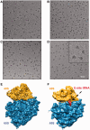
Ribosomes are key macromolecular protein synthesis machineries in the cell. Human ribosomes have so far not been studied to atomic resolution because of their particularly complex structure as compared with other eukaryotic or prokaryotic ribosomes, and they are difficult to prepare to high homogeneity, which is a key requisite for high-resolution structural work. We established a purification protocol for human 80S ribosomes isolated from HeLa cells that allows obtaining large quantities of homogenous samples as characterized by biophysical methods using analytical ultracentrifugation and multiangle laser light scattering. Samples prepared under different conditions were characterized by direct single particle imaging using cryo electron microscopy, which helped optimizing the preparation protocol. From a small data set, a 3D reconstruction at subnanometric resolution was obtained showing all prominent structural features of the human ribosome, and revealing a salt concentration dependence of the presence of the exit site tRNA, which we show is critical for obtaining crystals. With these well-characterized samples first human 80S ribosome crystals were obtained from several crystallization conditions in capillaries and sitting drops, which diffract to 26 Å resolution at cryo temperatures and for which the crystallographic parameters were determined, paving the way for future high-resolution work.
Figures


Similar articles
-
Cryo-EM structure and rRNA model of a translating eukaryotic 80S ribosome at 5.5-A resolution.Proc Natl Acad Sci U S A. 2010 Nov 16;107(46):19748-53. doi: 10.1073/pnas.1009999107. Epub 2010 Oct 27. Proc Natl Acad Sci U S A. 2010. PMID: 20980660 Free PMC article.
-
Structure of the human 80S ribosome.Nature. 2015 Apr 30;520(7549):640-5. doi: 10.1038/nature14427. Epub 2015 Apr 22. Nature. 2015. PMID: 25901680
-
Comprehensive molecular structure of the eukaryotic ribosome.Structure. 2009 Dec 9;17(12):1591-1604. doi: 10.1016/j.str.2009.09.015. Structure. 2009. PMID: 20004163 Free PMC article.
-
Crystal structure of the 80S yeast ribosome.Curr Opin Struct Biol. 2012 Dec;22(6):759-67. doi: 10.1016/j.sbi.2012.07.013. Epub 2012 Aug 8. Curr Opin Struct Biol. 2012. PMID: 22884264 Review.
-
Three-dimensional electron cryomicroscopy of ribosomes.Curr Protein Pept Sci. 2002 Feb;3(1):79-91. doi: 10.2174/1389203023380873. Curr Protein Pept Sci. 2002. PMID: 12370013 Review.
Cited by
-
MetAP-like Ebp1 occupies the human ribosomal tunnel exit and recruits flexible rRNA expansion segments.Nat Commun. 2020 Feb 7;11(1):776. doi: 10.1038/s41467-020-14603-7. Nat Commun. 2020. PMID: 32034140 Free PMC article.
-
The nucleoside analog clitocine is a potent and efficacious readthrough agent.RNA. 2017 Apr;23(4):567-577. doi: 10.1261/rna.060236.116. Epub 2017 Jan 17. RNA. 2017. PMID: 28096517 Free PMC article.
-
Visualization of chemical modifications in the human 80S ribosome structure.Nature. 2017 Nov 23;551(7681):472-477. doi: 10.1038/nature24482. Epub 2017 Nov 15. Nature. 2017. PMID: 29143818
-
Structural analysis of the SRP Alu domain from Plasmodium falciparum reveals a non-canonical open conformation.Commun Biol. 2021 May 20;4(1):600. doi: 10.1038/s42003-021-02132-y. Commun Biol. 2021. PMID: 34017052 Free PMC article.
-
Dynamic states of eIF6 and SDS variants modulate interactions with uL14 of the 60S ribosomal subunit.Nucleic Acids Res. 2023 Feb 28;51(4):1803-1822. doi: 10.1093/nar/gkac1266. Nucleic Acids Res. 2023. PMID: 36651285 Free PMC article.
References
-
- Dube P, Bacher G, Stark H, Mueller F, Zemlin F, van Heel M, Brimacombe R. Correlation of the expansion segments in mammalian rRNA with the fine structure of the 80 S ribosome; a cryoelectron microscopic reconstruction of the rabbit reticulocyte ribosome at 21 A resolution. J. Mol. Biol. 1998;279:403–421. - PubMed
-
- Chen IJ, Wang IA, Tai LR, Lin A. The role of expansion segment of human ribosomal protein L35 in nuclear entry, translation activity, and endoplasmic reticulum docking. Biochem. Cell Biol. 2008;86:271–277. - PubMed
-
- Klaholz BP. Molecular recognition and catalysis in translation termination complexes. Trends Biochem. Sci. 2011;36:282–292. - PubMed
Publication types
MeSH terms
Grants and funding
LinkOut - more resources
Full Text Sources
Other Literature Sources

