. 2014 Apr;42(7):4615-28.
doi: 10.1093/nar/gkt1391. Epub 2014 Jan 21.

Stepwise assembly of multiple Lin28 proteins on the terminal loop of let-7 miRNA precursors

Affiliations

Stepwise assembly of multiple Lin28 proteins on the terminal loop of let-7 miRNA precursors

Alexandre Desjardins et al. Nucleic Acids Res. 2014 Apr.

Abstract

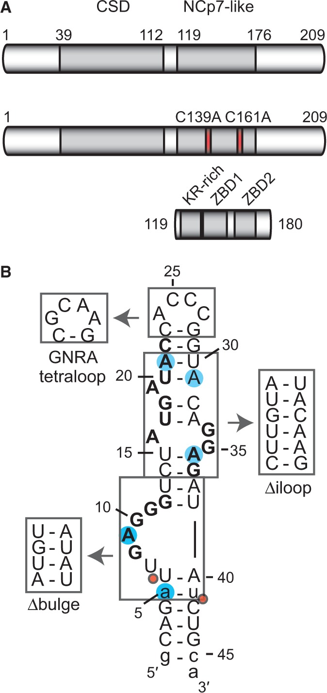
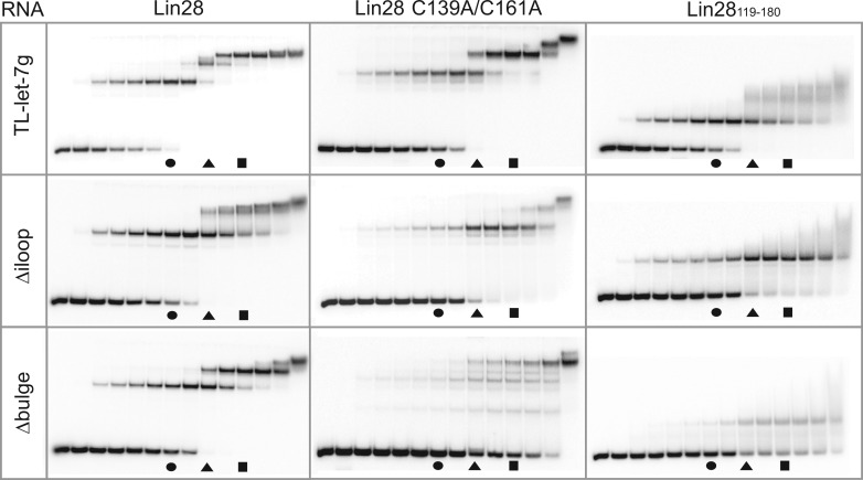
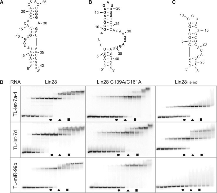
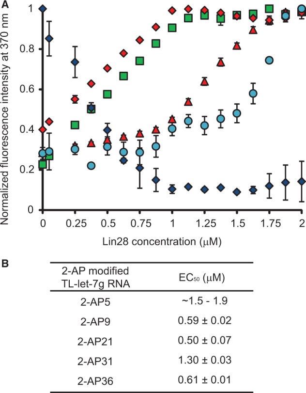
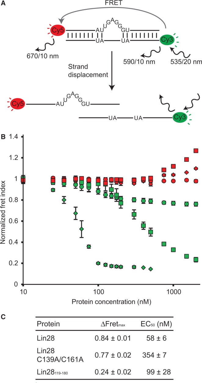
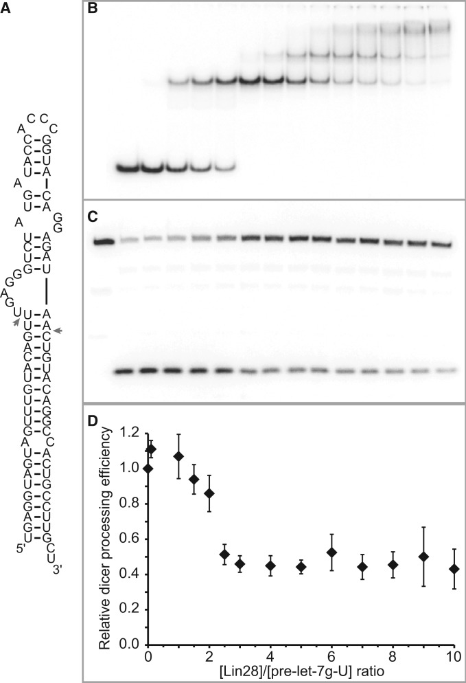
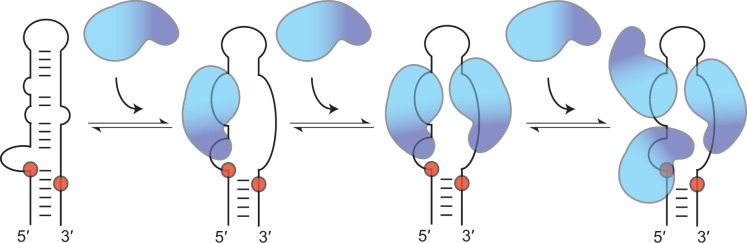
Lin28 inhibits the biogenesis of let-7 miRNAs through direct interactions with let-7 precursors. Previous studies have described seemingly inconsistent Lin28 binding sites on pre-let-7 RNAs. Here, we reconcile these data by examining the binding mechanism of Lin28 to the terminal loop of pre-let-7g (TL-let-7g) using biochemical and biophysical methods. First, we investigate Lin28 binding to TL-let-7g variants and short RNA fragments and identify three independent binding sites for Lin28 on TL-let-7g. We then determine that Lin28 assembles in a stepwise manner on TL-let-7g to form a stable 1:3 complex. We show that the cold-shock domain (CSD) of Lin28 is responsible for remodelling the terminal loop of TL-let-7g, whereas the NCp7-like domain facilitates the initial binding of Lin28 to TL-let-7g. This stable binding of multiple Lin28 molecules to the terminal loop of pre-let-7g extends to other precursors of the let-7 family, but not to other pre-miRNAs tested. We propose a model for stepwise assembly of the 1:1, 1:2 and 1:3 pre-let-7g/Lin28 complexes. Stepwise multimerization of Lin28 on pre-let-7 is required for maximum inhibition of Dicer cleavage for a least one member of the let-7 family and may be important for orchestrating the activity of the several factors that regulate let-7 biogenesis.


Figures

Figure 1.
Figure 1.
The Lin28 protein, TL-let-7g RNA and related sequences used in this study. (A) Schematic representation of the primary structures of Lin28 and related variants, Lin28 C139A/C161A and Lin28119–180. The grey boxes delineate sequences of known RNA-binding motifs: a CSD and an NCp7-like domain with a KR-rich region (residues 125–135) N-terminal to a pair of retroviral-type CCHC zinc-binding domains [ZBD1 (residues 137–154) and ZBD2 (residues 160–176); (19)]. Site-specific substitutions of Lin28 are shown in red. (B) Primary and proposed secondary structures of TL-let-7g. Nonnatural nucleotides are shown in lowercase, residues of previously identified Lin28-binding sites in bold characters, substitution sites for 2-AP are as blue shadows, regions that were replaced by alternative structural elements are boxed, and Dicer cleavage sites are indicated by red dots.
Figure 2.
Figure 2.
Stoichiometric binding assay by native gel electrophoresis for Lin28 binding to TL-let-7g RNA and effect of select RNA and protein variants on the stoichiometry of the complex. Each assay is performed with 0.5 µM RNA, including 10 pM 5′-[32P]-labelled RNA, and increasing concentrations of protein (0.00, 0.05, 0.25, 0.375, 0.50, 0.625, 0.75, 1.00, 1.25, 1.50, 1.75, 2.00, 2.50 and 5.00 µM). The gel lanes with RNA:protein ratios of 1:1.5, 1:2.5 and 1:3.5 are identified by a circle, a triangle and a square, respectively.
Figure 3.
Figure 3.
Stoichiometric binding assay by native gel electrophoresis for Lin28 binding to other pre-let-7 terminal loops. Sequences and proposed secondary structures of (A) TL-let-7a-1, (B) TL-let-7d and (C) TL-miR-99b with bold residues representing previously identified or predicted Lin28-binding sites (see text). Nonnatural nucleotides are shown in lowercase. (D) Stoichiometric binding assay of TL-let-7a-1, TL-let-7d and TL-miR-99b with Lin28, Lin28 C139A/C161A and Lin28119–180. Each assay is performed with 0.5 µM RNA, including 10 pM 5′-[32P]-labelled RNA, and increasing concentrations of protein (0.00, 0.05, 0.25, 0.375, 0.50, 0.625, 0.75, 1.00, 1.25, 1.50, 1.75, 2.00, 2.50 and 5.00 µM). The gel lanes with RNA:protein ratios of 1:1.5, 1:2.5 and 1:3.5 are identified by a circle, a triangle and a square, respectively.
Figure 4.
Figure 4.
Effect of Lin28 on the fluorescence intensity of TL-let-7g containing individual 2-AP modifications. (A) Normalized fluorescence intensity at 370 nm of 2-AP5 (cyan dots), 2-AP9 (green squares), 2-AP21 (blue diamonds), 2-AP31 (red triangles) and 2-AP36 (red diamonds) as a function of Lin28 concentration. Each titration point and the associated error bars are respectively the average and standard deviation from multiple experiments. (B) Reported EC50 values and their errors obtained from data in (A) by fitting the normalized fluorescence intensity at 370 nm with respect to Lin28 concentration using the dose–response equation.
Figure 5.
Figure 5.
Strand displacement assay monitored by FRET to test the RNA melting activity of Lin28. (A) Schematic of the RNA melting activity of Lin28 on the duplexbulge RNA, a Cy3/Cy5-labelled duplex. Relevant excitation and emission frequencies are indicated with associated slit widths. (B) Normalized Fret Index (FCy5/FCy3) of Cy3/Cy5-labelled RNA duplexes (25 nM) as a function of total protein concentration (10, 25, 35, 50, 60, 75, 100, 125, 150, 175, 200, 300, 400, 500, 750, 1000, 1500 and 2000 nM). Proteins were sequentially added to both the duplexbulge (Lin28, green diamonds; Lin28 C139A/C161A, green squares; and Lin28119–180, green circles) and the control duplexcomp (Lin28, red diamonds; Lin28 C139A/C161A, red squares; and Lin28119-180, red circles). (C) Values of ΔFRETmax and EC50 derived from results shown in (B).
Figure 6.
Figure 6.
Dicer processing assay of pre-let-7g-U. (A) Primary and proposed secondary structures of the pre-let-7g-U RNA. Dicer cleavage sites are identified with arrows. (B) Stoichiometric binding assay detected by native gel electrophoresis for Lin28 binding to pre-let-7g-U. Each assay is performed with 0.5 µM 5′-phosphorylated RNA, including 40 pM 5′-[32P]-labelled RNA and increasing concentration of Lin28 (0.00, 0.05, 0.50, 0.75, 1.25, 1.50, 2.00, 2.50, 3.00, 3.50, 4.00, 4.50 and 5.00 µM). (C) Dicer processing assay detected by denaturing gel electrophoresis of pre-let-7g-U under varying Lin28 concentrations. The first well contains 0.5 µM RNA without Lin28 and Dicer. The subsequent wells are for the Dicer assay performed under the same conditions as for the stoichiometric binding assay (in B), but with an additional 0.25 U of Dicer enzyme. (D) Relative Dicer processing efficiency plotted against Lin28/pre-let-7g-U concentration ratios (n = 2).
Figure 7.
Figure 7.
Schematic representation of the proposed model for the stepwise assembly of Lin28 on the terminal loop of pre-let-7g. In this model, only the RNA-binding domains of Lin28 are represented, with the NCp7-like domain in dark blue and the CSD in cyan. The first molecule of Lin28 initiates complex formation through interactions between its NCp7-like domain and the conserved 5′-GAGGG-3′ site at the 5′-bulge and between its CSD and the nearby 5′-AUGAUAC-3′ site. These interactions on the 5′-strand destabilize Watson–Crick base pairs within the terminal loop and exposes the 3′-strand, making it available for binding a second molecule of Lin28, with its NCp7-like domain targeting the conserved 5′-GGAG-3′ sequence and its CSD targeting an adjacent 5′-region. Binding of the third molecule of Lin28 to the high-affinity 5′-AUGAUAC-3′ site involves only its CSD and implicates relocating the CSD of the first molecule of Lin28. The order of assembly proposed for pre-let-7g may differ for precursors of other let-7 family members.

Similar articles

Cited by

References

    1. Bartel DP. MicroRNAs: target recognition and regulatory functions. Cell. 2009;136:215–233. - PMC - PubMed
    1. Kim VN, Han J, Siomi MC. Biogenesis of small RNAs in animals. Nat. Rev. Mol. Cell Biol. 2009;10:126–139. - PubMed
    1. Newman MA, Hammond SM. Emerging paradigms of regulated microRNA processing. Genes Dev. 2010;24:1086–1092. - PMC - PubMed
    1. Winter J, Diederichs S. MicroRNA biogenesis and cancer. Methods Mol. Biol. 2011;676:3–22. - PubMed
    1. Ambros V. MicroRNAs and developmental timing. Curr. Opin. Genet. Dev. 2011;21:511–517. - PMC - PubMed

Publication types