. 2014 Apr;42(7):4755-66.
doi: 10.1093/nar/gkt1389. Epub 2014 Jan 21.

Expanding the zinc-finger recombinase repertoire: directed evolution and mutational analysis of serine recombinase specificity determinants

Affiliations

Expanding the zinc-finger recombinase repertoire: directed evolution and mutational analysis of serine recombinase specificity determinants

Shannon J Sirk et al. Nucleic Acids Res. 2014 Apr.

Abstract

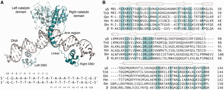
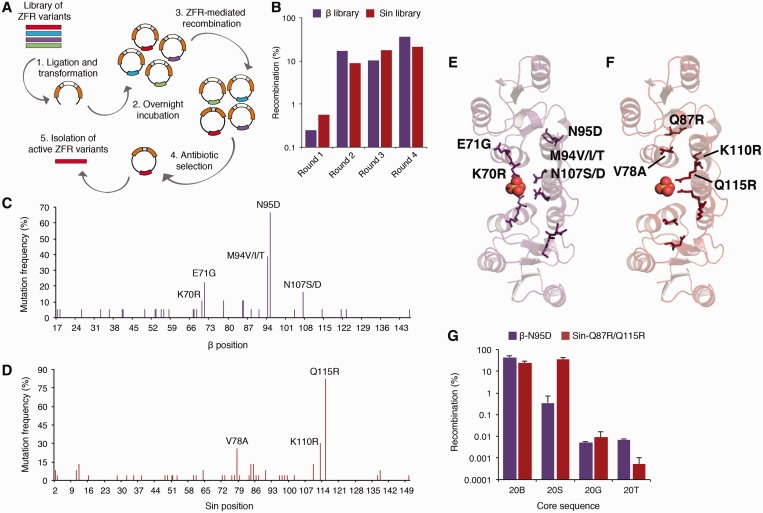
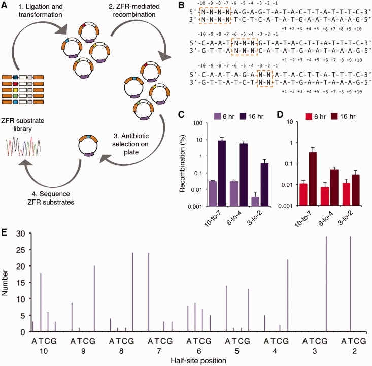
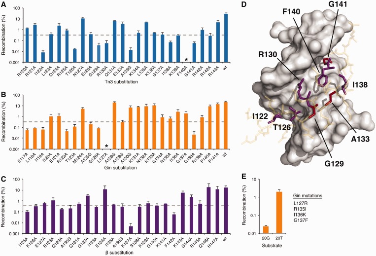
The serine recombinases are a diverse family of modular enzymes that promote high-fidelity DNA rearrangements between specific target sites. Replacement of their native DNA-binding domains with custom-designed Cys₂-His₂ zinc-finger proteins results in the creation of engineered zinc-finger recombinases (ZFRs) capable of achieving targeted genetic modifications. The flexibility afforded by zinc-finger domains enables the design of hybrid recombinases that recognize a wide variety of potential target sites; however, this technology remains constrained by the strict recognition specificities imposed by the ZFR catalytic domains. In particular, the ability to fully reprogram serine recombinase catalytic specificity has been impeded by conserved base requirements within each recombinase target site and an incomplete understanding of the factors governing DNA recognition. Here we describe an approach to complement the targeting capacity of ZFRs. Using directed evolution, we isolated mutants of the β and Sin recombinases that specifically recognize target sites previously outside the scope of ZFRs. Additionally, we developed a genetic screen to determine the specific base requirements for site-specific recombination and showed that specificity profiling enables the discovery of unique genomic ZFR substrates. Finally, we conducted an extensive and family-wide mutational analysis of the serine recombinase DNA-binding arm region and uncovered a diverse network of residues that confer target specificity. These results demonstrate that the ZFR repertoire is extensible and highlights the potential of ZFRs as a class of flexible tools for targeted genome engineering.


Figures

Figure 1.
Figure 1.
Overview of the small serine recombinases. (A) (Top) Crystal structure of the γδ resolvase dimer bound to target DNA (PDB ID: 1GDT) (20). ‘Left’ and ‘right’ recombinase monomers are colored light and dark teal, respectively. DBD indicates native DNA-binding domain. Linker and arm region are labeled for the ‘right’ recombinase monomer only. (Bottom) Core sequence recognized by the γδ resolvase catalytic domain. Base positions are indicated. (B) Sequence alignment of six of the most comprehensively characterized serine recombinase catalytic domains. Conserved residues are highlighted light teal. The α-helical and β-sheet secondary structural elements are denoted above the alignment as cylinders and arrows, respectively.
Figure 2.
Figure 2.
Directed evolution of enhanced β and Sin catalytic domains. (A) Schematic representation illustrating the split gene reassembly selection strategy. ZFR variants are shown in various colors; β-lactamase gene is in orange and GFPuv gene is in white. (B) Selection of β and Sin variants that recombine minimal core sites from the six and resH recombination sites, respectively. (C, D) Frequency and position of the mutations that activate the (C) β and (D) Sin catalytic domains. Highly recurrent mutations are indicated. (E, F) Crystal structure of the activated Sin-Q115R tetramer; view of dimer interface from above the N-terminus of the E helix (PDB ID: 3PKZ) (51). Highly recurrent (E) β and (F) Sin mutations shown as sticks and mapped onto the rotated Sin dimer, residues labeled on upper monomer only. Sulfate ion shown as spheres. (G) Recombination activity of β-N95D and Sin-Q87R/Q115R on the 20B, 20S, 20G and 20T core sequences. Recombination was determined by split gene reassembly. Error bars indicate standard deviation (n = 3).
Figure 3.
Figure 3.
Specificity of the β-N95D catalytic domain. (A) Schematic representation illustrating the genetic screen used to profile recombinase specificity. Recombinase substrate library shown in various colors; ZFR gene is in purple, β-lactamase gene is in orange and GFPuv gene is in white. (B) Randomization strategy used for specificity profiling. Randomized bases are boxed. Note that only ‘left’ half-site of the upstream ZFR target site contained base substitutions. (C and D) Recombination by (C) β-N95D and (D) Sin-Q87R/Q115R for each 20B and 20S core site library, respectively, at 6 and 16 h. (E) Number of selected base sequences (out of 30) at each position within the 20B half-site. Thirty clones were sequenced from each 6-h library output. Recombination was determined by split gene reassembly. Error bars indicate standard deviation (n = 3).
Figure 4.
Figure 4.
Alanine-scanning mutagenesis of the serine recombinase arm region. (A–C) Recombination activity of mutant (A) Tn3, (B) Gin and (C) β catalytic domains on their native and minimal DNA targets. Asterisk indicates <0.0001% recombination. Dotted lines indicate threshold below which mutants were considered non-functional. (D) Crystal structure of the γδ resolvase arm region (sticks) in contact with substrate DNA (gray surface). Conserved and variable residues important for recombination are shown in red and purple, respectively. Inert residues are shown in yellow (PDB ID: 1GDT) (20). (E) Recombination by a Gin chimera substituted with residues predicted to impart specificity onto the 20T core site. Recombination was determined by split gene reassembly. Error bars indicate standard deviation (n = 3).

Similar articles

Cited by

References

    1. Gaj T, Gersbach CA, Barbas CF., III ZFN, TALEN, and CRISPR/Cas-based methods for genome engineering. Trends Biotechnol. 2013;31:397–405. - PMC - PubMed
    1. Bibikova M, Beumer K, Trautman JK, Carroll D. Enhancing gene targeting with designed zinc finger nucleases. Science. 2003;300:764. - PubMed
    1. Perez EE, Wang J, Miller JC, Jouvenot Y, Kim KA, Liu O, Wang N, Lee G, Bartsevich VV, Lee YL, et al. Establishment of HIV-1 resistance in CD4+ T cells by genome editing using zinc-finger nucleases. Nat. Biotechnol. 2008;26:808–816. - PMC - PubMed
    1. Gaj T, Guo J, Kato Y, Sirk SJ, Barbas CF., III Targeted gene knockout by direct delivery of zinc-finger nuclease proteins. Nat. Methods. 2012;9:805–807. - PMC - PubMed
    1. Miller JC, Tan S, Qiao G, Barlow KA, Wang J, Xia DF, Meng X, Paschon DE, Leung E, Hinkley SJ, et al. A TALE nuclease architecture for efficient genome editing. Nat. Biotechnol. 2011;29:143–148. - PubMed

Publication types