

Synergies of phosphatidylserine and protein disulfide isomerase in tissue factor activation
- PMID: 24452853
- PMCID: PMC4270292
- DOI: 10.1160/TH13-09-0802
Synergies of phosphatidylserine and protein disulfide isomerase in tissue factor activation
Abstract
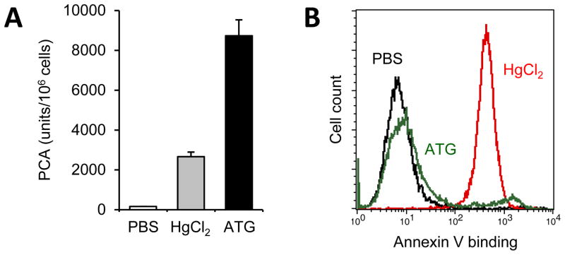
Tissue factor (TF), the cellular receptor and cofactor for factor VII/VIIa, initiates haemostasis and thrombosis. Initial tissue distribution studies suggested that TF was sequestered from the circulation and only present at perivascular sites. However, there is now clear evidence that TF also exists as a blood-borne form with critical contributions not only to arterial thrombosis following plaque rupture and to venous thrombosis following endothelial perturbation, but also to various other clotting abnormalities associated with trauma, infection, or cancer. Because thrombin generation, fibrin deposition, and platelet aggregation in the contexts of haemostasis, thrombosis, and pathogen defence frequently occur without TF de novo synthesis, considerable efforts are still directed to understanding the molecular events underlying the conversion of predominantly non-coagulant or cryptic TF on the surface of haematopoietic cells to a highly procoagulant molecule following cellular injury or stimulation. This article will review some of the still controversial mechanisms implicated in cellular TF activation or decryption with particular focus on the coordinated effects of outer leaflet phosphatidylserine exposure and thiol-disulfide exchange pathways involving protein disulfide isomerase (PDI). In this regard, our recent findings of ATP-triggered stimulation of the purinergic P2X7 receptor on myeloid and smooth muscle cells resulting in potent TF activation and shedding of procoagulant microparticles as well as of rapid monocyte TF decryption following antithymocyte globulin-dependent membrane complement fixation have delineated specific PDI-dependent pathways of cellular TF activation and thus illustrated additional and novel links in the coupling of inflammation and coagulation.
Keywords: Tissue factor; phosphatidylserine; protein disulfide isomerase; thiol-disulfide exchange; thrombosis.
Conflict of interest statement
The authors declare no competing financial interests.
Figures


Comment in
-
Activators, therapeutics and immunity-related aspects of thrombosis.Thromb Haemost. 2014 Apr 1;111(4):568-9. doi: 10.1160/TH14-03-0203. Epub 2014 Mar 20. Thromb Haemost. 2014. PMID: 24651966 No abstract available.
References
Publication types
MeSH terms
Substances
Grants and funding
LinkOut - more resources
Full Text Sources
Other Literature Sources
Medical
Miscellaneous
